Unusual brachial plexus lesion: Hematoma masquerading as a peripheral nerve sheath tumor
- PMID: 26904368
- PMCID: PMC4743268
- DOI: 10.4103/2152-7806.174889
Unusual brachial plexus lesion: Hematoma masquerading as a peripheral nerve sheath tumor
Abstract
Background: Malignant peripheral nerve sheath tumors (MPNSTs) of the brachial plexus have unique radiographic and clinical findings. Patients often present with progressive upper extremity paresthesias, weakness, and pain. On magnetic resonance (MR) imaging, lesions are isointense on T1-weighted and hyperintense on T2-weighted sequences, while also demonstrating marked enhancement on MR studies with gadolinium diethylenetriamine pentaacetic acid. On the basis of their characteristic MR imaging features and rapid clinical progression, two brachial plexus lesions proved to be organizing hematomas rather than MPNST.
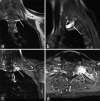
Methods: A 51-year-old male and a 31-year-old female were both assessed for persistent and worsened left-sided upper extremity pain, paresthesias, and weakness. In both cases, the MR imaging of the brachial plexus demonstrated an extraspinal enhancing lesion located within the left C7-T1 neuroforamina.
Results: Although the clinical and radiographic MR features for these 2 patients were consistent with MPNSTs, both lesions proved to be benign organizing hematomas.
Conclusions: These two case studies emphasize that brachial plexus hematomas may mimic MPNSTs on MR studies. Accurate diagnosis of these lesions is critical for determining the appropriate management options and treatment plans. Delaying the treatment of a highly aggressive nerve sheath tumor can have devastating consequences, whereas many hematomas resolve without surgery. Therefore, if the patient has stable findings on neurological examination and a history of trauma, surgical intervention may be delayed in favor of repeat MR imaging in 2-3 months to re-evaluate the size of the mass.
Keywords: Brachial plexus; hematoma; malignant peripheral nerve sheath tumor; nerve sheath tumor; schwannoma.
Figures


Similar articles
-
Brachial plexus peripheral nerve sheath tumors (PNSTs): clinical and surgical management in the pediatric population.Childs Nerv Syst. 2024 Nov;40(11):3789-3800. doi: 10.1007/s00381-024-06509-2. Epub 2024 Jun 28. Childs Nerv Syst. 2024. PMID: 38940956
-
Magnetic resonance imaging characteristics of peripheral nerve sheath tumors of the canine brachial plexus in 18 dogs.Vet Radiol Ultrasound. 2007 Jan-Feb;48(1):1-7. doi: 10.1111/j.1740-8261.2007.00195.x. Vet Radiol Ultrasound. 2007. PMID: 17236352
-
Malignant peripheral nerve sheath tumor: the utility of fascicular biopsy and teased fiber studies.J Clin Neuromuscul Dis. 2012 Sep;14(1):28-33. doi: 10.1097/CND.0b013e318260b396. J Clin Neuromuscul Dis. 2012. PMID: 22922579
-
MR imaging of the brachial plexus: current imaging sequences, normal findings, and findings in a spectrum of focal lesions with MR-pathologic correlation.Curr Probl Diagn Radiol. 2003 Mar-Apr;32(2):88-101. doi: 10.1067/mdr.2003.12007. Curr Probl Diagn Radiol. 2003. PMID: 12658265 Review.
-
Imaging tumours of the brachial plexus.Skeletal Radiol. 2003 Jul;32(7):375-87. doi: 10.1007/s00256-003-0618-0. Epub 2003 Mar 20. Skeletal Radiol. 2003. PMID: 12851788 Review.
Cited by
-
Tumor-like disorder of the brachial plexus region in a patient with hemophilia: A case report.World J Clin Cases. 2022 Jun 16;10(17):5910-5915. doi: 10.12998/wjcc.v10.i17.5910. World J Clin Cases. 2022. PMID: 35979120 Free PMC article.
References
-
- Ducatman BS, Scheithauer BW, Piepgras DG, Reiman HM, Ilstrup DM. Malignant peripheral nerve sheath tumors. A clinicopathologic study of 120 cases. Cancer. 1986;57:2006–21. - PubMed
-
- Enzinger F, Weiss S. St. Louis, MO: CV Mosby; 1983. Soft Tissue Tumors.
-
- Ganju A, Roosen N, Kline DG, Tiel RL. Outcomes in a consecutive series of 111 surgically treated plexal tumors: A review of the experience at the Louisiana State University Health Sciences Center. J Neurosurg. 2001;95:51–60. - PubMed
-
- Niimi R, Matsumine A, Kusuzaki K, Okamura A, Matsubara T, Uchida A, et al. Soft-tissue sarcoma mimicking large haematoma: A report of two cases and review of the literature. J Orthop Surg (Hong Kong) 2006;14:90–5. - PubMed
-
- Wasa J, Nishida Y, Tsukushi S, Shido Y, Sugiura H, Nakashima H, et al. MRI features in the differentiation of malignant peripheral nerve sheath tumors and neurofibromas. AJR Am J Roentgenol. 2010;194:1568–74. - PubMed
LinkOut - more resources
Full Text Sources
Other Literature Sources
Miscellaneous

