

Detrusor sphincter dyssynergia: a review of physiology, diagnosis, and treatment strategies
- PMID: 26904418
- PMCID: PMC4739973
- DOI: 10.3978/j.issn.2223-4683.2016.01.08
Detrusor sphincter dyssynergia: a review of physiology, diagnosis, and treatment strategies
Abstract
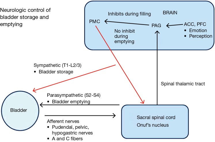
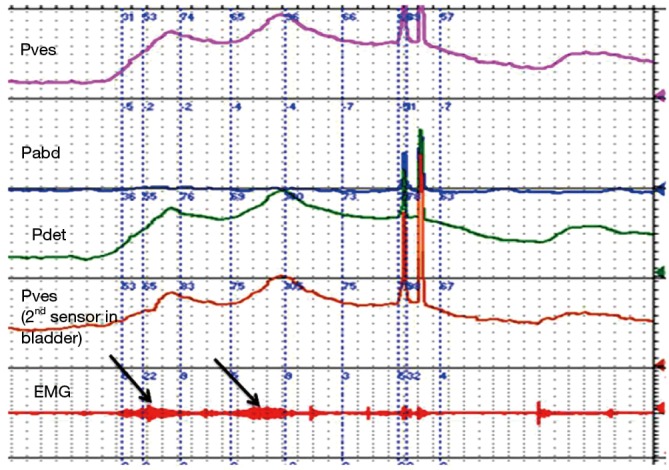
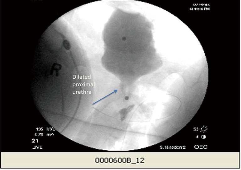
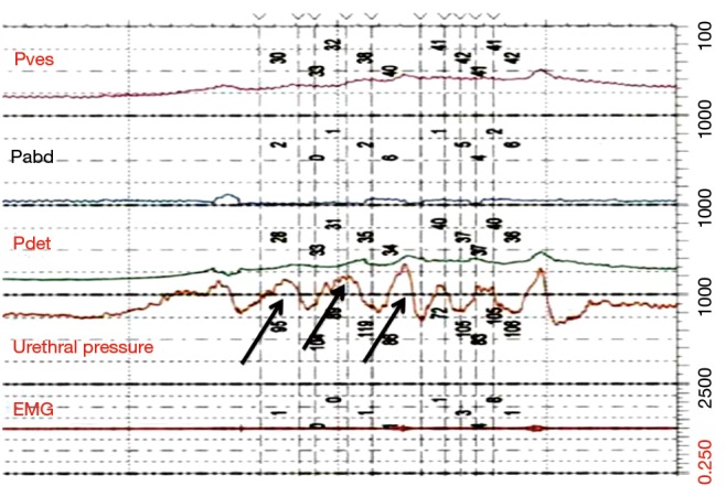
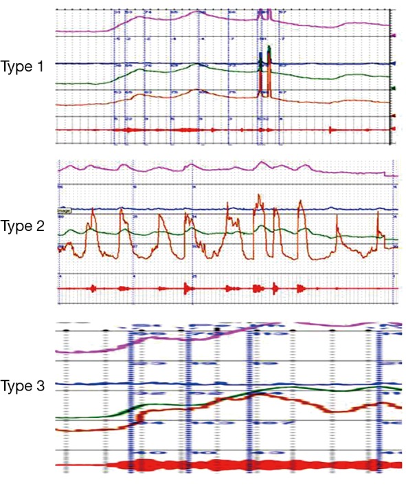
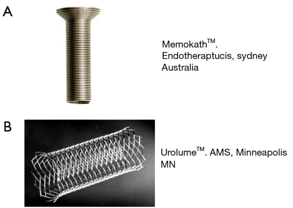
Detrusor sphincter dyssynergia (DSD) is the urodynamic description of bladder outlet obstruction from detrusor muscle contraction with concomitant involuntary urethral sphincter activation. DSD is associated with neurologic conditions such as spinal cord injury, multiple sclerosis, and spina bifida and some of these neurogenic bladder patients with DSD may be at risk for autonomic dysreflexia, recurrent urinary tract infections, or upper tract compromise if the condition is not followed and treated appropriately. It is diagnosed most commonly during the voiding phase of urodynamic studies using EMG recordings and voiding cystourethrograms, although urethral pressure monitoring could also potentially be used. DSD can be sub-classified as either continuous or intermittent, although adoption of this terminology is not widespread. There are few validated oral pharmacologic treatment options for this condition but transurethral botulinum toxin injection have shown temporary efficacy in reducing bladder outlet obstruction. Urinary sphincterotomy has also demonstrated reproducible long term benefits in several studies, but the morbidity associated with this procedure can be high.
Keywords: Detrusor sphincter dyssynergia (DSD); external urinary sphincter (EUS); neurogenic bladder; urodynamics.
Conflict of interest statement
Figures
References
Publication types
LinkOut - more resources
Full Text Sources