

Developmental Structural Tooth Defects in Dogs - Experience From Veterinary Dental Referral Practice and Review of the Literature
- PMID: 26904551
- PMCID: PMC4744861
- DOI: 10.3389/fvets.2016.00009
Developmental Structural Tooth Defects in Dogs - Experience From Veterinary Dental Referral Practice and Review of the Literature
Abstract
Developmental tooth abnormalities in dogs are uncommon in general veterinary practice but understanding thereof is important for optimal management in order to maintain masticatory function through preservation of the dentition. The purpose of this review is to discuss clinical abnormalities of the enamel and general anatomy of dog teeth encountered in veterinary dental referral practice and described in the literature. More than 900 referral cases are seen annually between the two referral practices. The basis of the pathogenesis, resultant clinical appearance, and the principles of management for each anomaly will be described. Future research should be aimed toward a more detailed analysis of these conditions so rarely described in the literature.
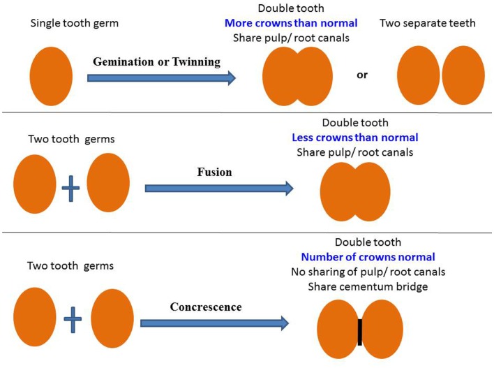
Keywords: concrescence; enamel hypoplasia; fusion; gemination; odontogenesis; tooth abnormalities.
Figures

Similar articles
-
Clinical diagnosis of enamel defects: pitfalls and practical guidelines.Int Dent J. 1997 Jun;47(3):173-82. doi: 10.1002/j.1875-595x.1997.tb00783.x. Int Dent J. 1997. PMID: 9448804 Review.
-
[Veterinary dentistry (3). Development, anatomy and function of teeth in the dog].Tijdschr Diergeneeskd. 1991 Nov 15;116(22):1107-21. Tijdschr Diergeneeskd. 1991. PMID: 1957295 Dutch.
-
Coincidence of Fusion and Concrescence in Mandibular Deciduous Incisors: A Case Report.J Contemp Dent Pract. 2019 Dec 1;20(12):1466-1469. J Contemp Dent Pract. 2019. PMID: 32381851
-
Developmental enamel defects in the primary dentition: aetiology and clinical management.Aust Dent J. 2013 Jun;58(2):133-40; quiz 266. doi: 10.1111/adj.12039. Epub 2013 May 5. Aust Dent J. 2013. PMID: 23713631 Review.
-
Rare dental developmental disturbance in primary and permanent teeth following trauma prior to tooth eruption: Case report.Dent Traumatol. 2020 Feb;36(1):79-83. doi: 10.1111/edt.12500. Epub 2019 Nov 20. Dent Traumatol. 2020. PMID: 31234235
Cited by
-
An autosomal recessive mutation in SCL24A4 causing enamel hypoplasia in Samoyed and its relationship to breed-wide genetic diversity.Canine Genet Epidemiol. 2017 Nov 22;4:11. doi: 10.1186/s40575-017-0049-1. eCollection 2017. Canine Genet Epidemiol. 2017. PMID: 29201383 Free PMC article.
-
Mandibular Carnassial Tooth Malformations in 6 Dogs-Micro-Computed Tomography and Histology Findings.Front Vet Sci. 2019 Dec 17;6:464. doi: 10.3389/fvets.2019.00464. eCollection 2019. Front Vet Sci. 2019. PMID: 31956654 Free PMC article.
-
An update on oral manifestations of systemic disorders in dogs and cats.Front Vet Sci. 2025 Jan 6;11:1511971. doi: 10.3389/fvets.2024.1511971. eCollection 2024. Front Vet Sci. 2025. PMID: 39834923 Free PMC article. Review.
-
Advancements in nanohydroxyapatite: synthesis, biomedical applications and composite developments.Regen Biomater. 2024 Nov 5;12:rbae129. doi: 10.1093/rb/rbae129. eCollection 2025. Regen Biomater. 2024. PMID: 39776858 Free PMC article. Review.
-
Incisor Disorders of Merino Sheep (Ovis aries).J Vet Dent. 2025 Sep;42(5):331-348. doi: 10.1177/08987564251339058. Epub 2025 May 8. J Vet Dent. 2025. PMID: 40340664 Free PMC article.
References
-
- Pavlica Z, Erjavec V, Petelin M. Teeth abnormalities in the dog. Acta Vet Brno (2001) 70:65–72.10.2754/avb200170010065 - DOI
Publication types
LinkOut - more resources
Full Text Sources
Other Literature Sources