. 2016 Feb 24:7:10809.
doi: 10.1038/ncomms10809.

Human oocyte developmental potential is predicted by mechanical properties within hours after fertilization

Affiliations

Human oocyte developmental potential is predicted by mechanical properties within hours after fertilization

Livia Z Yanez et al. Nat Commun. .

Abstract

The causes of embryonic arrest during pre-implantation development are poorly understood. Attempts to correlate patterns of oocyte gene expression with successful embryo development have been hampered by the lack of reliable and nondestructive predictors of viability at such an early stage. Here we report that zygote viscoelastic properties can predict blastocyst formation in humans and mice within hours after fertilization, with >90% precision, 95% specificity and 75% sensitivity. We demonstrate that there are significant differences between the transcriptomes of viable and non-viable zygotes, especially in expression of genes important for oocyte maturation. In addition, we show that low-quality oocytes may undergo insufficient cortical granule release and zona-hardening, causing altered mechanics after fertilization. Our results suggest that embryo potential is largely determined by the quality and maturation of the oocyte before fertilization, and can be predicted through a minimally invasive mechanical measurement at the zygote stage.


Figures
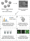
Figure 1
Figure 1. Overview of experimental design.
(a) We measured mechanical properties of 282 mouse and 89 human zygotes, and found the combination most predictive of viability, defined as survival to the blastocyst stage. (b) We then measured live birth rates in mice that received either embryos that were predicted to be viable based on mechanics (n=55), predicted non-viable based on mechanics (n=55) or randomly chosen as a control (n=55). (c) We conducted single-cell RNA-seq on 17 human zygotes and found differences in transcriptomes between those predicted to be viable and non-viable based on mechanics. (d) We investigated differences in cortical granule release between 30 embryos predicted to be viable and 37 embryos predicted to be non-viable based on mechanics. Scale bar, 40 μm.
Figure 2
Figure 2. Design of the measurement system and differences in the mechanical parameters between viable and non-viable embryos.
(a) The measurement technique and (b) the mechanical model used to extract mechanical parameters. (c) A typical trace of the pressure applied to the embryo through the micropipette (red line) and the response of the embryo to the pressure as it is aspirated into the micropipette (blue line). (d) Box-and-whisker plots (box shows median, edges are at first and third quartiles, so total box height is interquartile range (IQR), lower whisker extends from lower edge to lowest value within 1.5*IQR of the edge, upper whisker extends from upper edge to highest value within 1.5*IQR of the edge), with scatterplots overlaid of the mechanical parameters of human zygotes (n=89). Parameters of the viable zygotes (n=31) appear more tightly clustered than those of non-viable zygotes (n=58). (e) Three-dimensional scatter plot of three mechanical parameters showing that viable human zygotes (n=31) cluster tightly in one region, with non-viable zygotes (n=58) scattered around them. (f) Example images of some of the human zygotes with similar morphological scores that are predicted to be viable or non-viable based on mechanics. (g) The mechanical parameters were also predictive of live birth in mouse, and enabled us to improve live birth rates (P=0.01, χ2-test) compared with a group of control embryos (n=55 in each group, over a total of four replicates). *P<0.05, ***P<0.001. Error bars represent s.d. Scale bar, 60 μm.
Figure 3
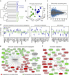
Figure 3. Results of RNA-seq on eight predicted viable and nine predicted non-viable human zygotes (predicted based on mechanics).
(a) Hierarchical clustering of gene expression values show that zygotes cluster by viability. N=8 viable and n=9 non-viable zygotes were used over a total of two replicates. (b) 3D principal component plot also shows that zygotes cluster by viability. (c) Using the edgeR package in R, we found 2,522 genes with statistically significant (q-value<0.01) differences in expression between viable and non-viable embryos (blue circles), most of which had a log-fold change of at least 0.5. (d) We found differences in many gene categories important in oocyte maturation and fertilization. Box plots containing the median and IQR of expression of particularly interesting genes are shown here. Whiskers extend up to 1.5 times the IQR from the box edges as in Fig. 2d. (e) IPA predicted a significant decrease in cell cycle checkpoint control and chromosome segregation in non-viable embryos (P<0.01). (f) A network of genes predicted to interact with each other (P<0.01), which are involved in chromatin modification, a process important for the oocyte-to-embryo transition.
Figure 4
Figure 4. Cortical granule staining reveals differences between fertilization between predicted viable and non-viable embryos based on mechanics.
(a) List of some of the genes important for fertilization identified as differentially expressed in RNA-seq results. (b) Representative images of mouse embryos stained for cortical granules. (c) Non-viable embryos have higher signal from cortical granules, regardless of whether they are too soft (n=24, P<10e−6, Wilcoxon rank sum test) or too stiff (n=13, P<10e−5, Wilcoxon rank sum test) compared with viable embryos (n=30). ***P<0.001. (d) Blocking of IP3 receptor in oocytes (n=32) before fertilization resulted in slightly softer mechanics after fertilization compared with control embryos (n=19), and absence of very stiff embryos. (e) Stiffest quartile of antibody-injected embryos showed lowered stiffness after IVF compared with stiffest quartile of control-injected embryos (P=0.02, Wilcoxon rank sum test), and increased brightness (arbitrary units) from unreleased cortical granules compared with control embryos with similar mechanical properties (P<0.01, Wilcoxon rank sum test). (f) Oocytes become less stiff (P<10e−7, Wilcoxon rank sum test) over the course of maturation from the GV (n=35) to MI (n=49) to MII (n=65) stages. Error bars represent s.d.. Scale bar, 25 μm.
Figure 5
Figure 5. Model for how embryo fate is determined and why it is detectable mechanically.
Some oocytes fail to achieve optimal maturation because of external stress during maturation, or inherent poor quality. At the time of fertilization, these oocytes (blue cytoplasm) may be overly stiff and still immature, or they could fail to release cortical granules properly and be overly soft after fertilization. Optimally matured oocytes (green cytoplasm) will release cortical granules into the perivitelline space, undergo the appropriate changes in mechanical properties and go on to develop successfully.

References

    1. Mtango N. R., Potireddy S. & Latham K. E. Oocyte quality and maternal control of development. Int. Rev. Cell Mol. Biol. 268, 223–290 (2008). - PubMed
    1. Stitzel M. L. & Seydoux G. Regulation of the oocyte-to-zygote transition. Science 316, 407–408 (2007). - PubMed
    1. Li L., Zheng P. & Dean J. Maternal control of early mouse development. Development 137, 859–870 (2010). - PMC - PubMed
    1. Bouniol C., Nguyen E. & Debey P. Endogenous transcription occurs at the 1-cell stage in the mouse embryo. Exp. Cell Res. 218, 57–62 (1995). - PubMed
    1. Dobson A. T. et al.. The unique transcriptome through day 3 of human preimplantation development. Hum. Mol. Genet. 13, 1461–1470 (2004). - PubMed

Publication types