. 2016 Mar 5;129(5):511-7.
doi: 10.4103/0366-6999.176988.

Inverted Internal Limiting Membrane Flap Technique for Repair of Large Macular Holes: A Short-term Follow-up of Anatomical and Functional Outcomes

Affiliations

Inverted Internal Limiting Membrane Flap Technique for Repair of Large Macular Holes: A Short-term Follow-up of Anatomical and Functional Outcomes

Zhe Chen et al. Chin Med J (Engl). .

Abstract

Background: Inverted internal limiting membrane (ILM) flap technique has recently been reported in a limited number of studies as an effective surgical technique for the management of large macular holes (MHs) with fair MH closure rates as well as gains in visual acuity. In the current study, longitudinal changes in multi-focal electroretinogram (mfERG) responses, best-corrected visual acuity (BCVA) and spectral-domain optical coherence tomography (SD-OCT) were evaluated in eyes with large MHs managed by this technique.

Methods: A prospective noncontrolled interventional study of eight patients (eight eyes) with large MHs (minimum diameter >400 μm) was conducted. All MHs were treated with pars plana vitrectomy and indocyanine green-assisted inverted ILM flap technique. SD-OCT images were used to assess the anatomical outcomes of surgery while BCVA and mfERG were used to evaluate the functional outcomes during a 3-month follow-up.

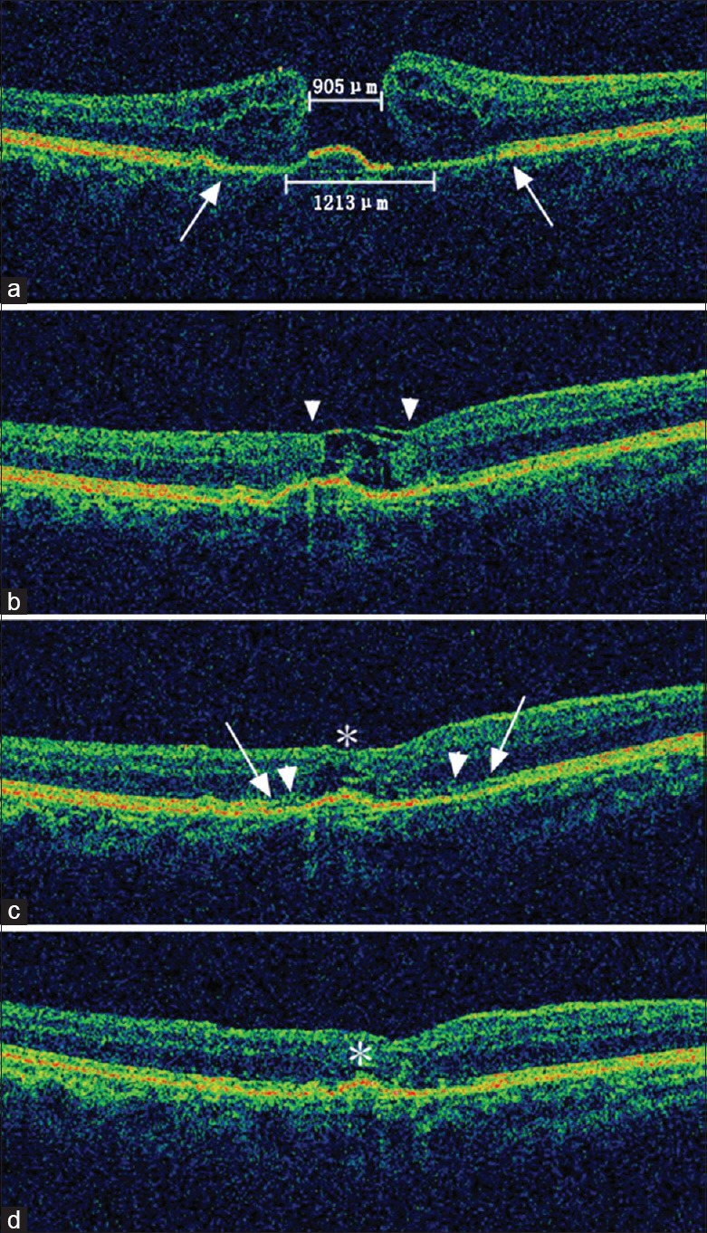
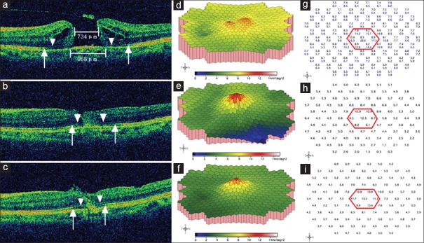
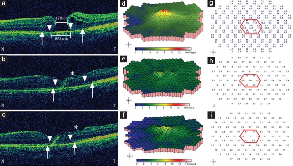
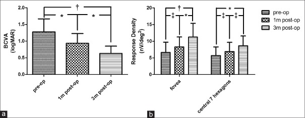
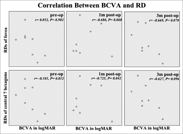
Results: All patients underwent successful intended manipulation and translocation of the ILM flap without flap dislocation and achieved complete anatomical closure. Partial microstructural reconstruction, demonstrated on SD-OCT as restoration of the external limiting membrane and the ellipsoid zone, was observed in all cases as early as 1 month after surgery. Functionally, as compared to baseline, all patients showed improvements in BCVA and all but one in mfERG response during follow-up. However, Pearson's test revealed no significant correlations between BCVA and mfERG responses of the fovea and of the macular area at each evaluation time point.

Conclusions: Inverted ILM flap technique appears to be a safe and effective approach for the management of large idiopathic MHs with favorable short-term anatomical and functional results. Postoperative reconstruction of the microstructure generally shows good consistency with improvements in both BCVA and mfERG response, of which the latter might be a supplement for the former in postoperative functional follow-up.


Figures

Figure 1
Figure 1
Spectral-domain optical coherence tomography images of the microstructural reconstruction process of a large macular hole in a 66-year-old female patient. (a) The preoperative minimum and base diameter of the macular hole was 905 μm and 1213 μm, respectively. Note the cystoid edema of the elevated margin and the disrupted external limiting membrane/ellipsoid zone (between the two arrows). The initial best corrected visual acuity was 0.08 (20/250). (b) Closure of the macular hole was observed 2 weeks after surgery, although the tissue filling the macular hole appeared to be sheets of inverted internal limiting membrane (flap closure, arrowheads). The best corrected visual acuity improved to 0.10 (20/200). (c) One month after surgery, the inner layers of the neurosensory retina thickened along the internal limiting membrane scaffold, leaving only a small defect of the outer retina (asterisk). The leading edge of the reconstructing external limiting membrane (arrowheads) and the ellipsoid zone (arrows) were both growing towards the center of the macular hole compared with the original sites before surgery. The best corrected visual acuity at this time was 0.16 (20/125). (d) Three months after surgery, the macular contour appeared further normalized, and the neurosensory retinal tissue above the fovea further thickened, leaving only a cleft (asterisk). Although not fully recovered at this stage, the external limiting membrane and the ellipsoid zone appeared more regular and clear. The best corrected visual acuity was 0.32 (20/63).
Figure 2
Figure 2
Representative spectral-domain optical coherence tomography and multi-focal electroretinogram changes of a 53-year-old female patient preoperative minimum and base diameters were 734 μm and 965 μm, respectively; defects of the external limiting membrane and the ellipsoid zone were indicated by arrow heads and arrows, respectively. Cystoid edema and elevation of the margin were demonstrated (a). On three-dimensional plot of multi-focal electroretinogram, the foveal and perifoveal area showed two separate small “peaks” (d), which reflected marked depression of the macular area and poor fixation due to low central vision. The response density of the central seven hexagons in this patient (g, the black numbers within the red hexagon) were much lower than a normal subject (g, the blue numbers). The initial best corrected visual acuity was 0.04 (20/500). One month after surgery, the macular hole closed, with partial restoration of both the external limiting membrane and the ellipsoid zone (b). The corresponding multi-focal electroretinogram showed a confluent blunt single peak (e), and the average retinal response density of the central seven hexagons and the fovea had increased from preoperative 8.5 and 8.8 nV/deg2 to 9.5 and 12.3 nV/deg2, respectively (h). The best corrected visual acuity was 0.13 (20/160) at this visit. Three months postoperatively, a further inward reconstruction of the external limiting membrane and the ellipsoid zone were observed (c). On three-dimensional plot of multi-focal electroretinogram, the peak became more sharp, with higher elevated immediate surrounding area (f); the corresponding average retinal response density of the central seven hexagons and the fovea had increased to 11.1 and 12.5 nV/deg2, respectively (i). The 3-month postoperative best corrected visual acuity was 0.20 (20/100).
Figure 3
Figure 3
Representative spectral-domain optical coherence tomography and multi-focal electroretinogram changes in a 67-year-old female patient preoperative minimum and base diameters were 478 μm and 854 μm, respectively; defects of the external limiting membrane were indicated by arrow heads and the ellipsoid zone by arrows. Letter “N” and “T” represent “nasal” and “temporal” side of the macula, respectively. At baseline, on spectral-domain optical coherence tomography, cystoid edema was more prominent on the temporal side of the macular hole (a), and there was a single small peak on three-dimensional plot of multi-focal electroretinogram (d) and the average retinal response density of the central seven hexagons and the fovea (g, the black numbers within the red hexagon) were 8.4 and 10.1 nV/deg2, respectively; the initial best corrected visual acuity was 0.02 (20/1000). One-month postoperatively, the macular hole had closed, but the defects of the external limiting membrane and the ellipsoid zone were larger than baseline levels, especially on the temporal side; note also the rugged inner surface of the temporal macula (asterisk) (b); the central peak on the three-dimensional plot of multi-focal electroretinogram was further depressed, (e) with average retinal response density of the central seven hexagons and the fovea decreased to 5.0 and 6.0 nV/deg2, respectively (h); however, the best-corrected visual acuity still increased to 0.05 (20/400). Three months after surgery, defects of the external limiting membrane and the ellipsoid zone were partially restored although the inner surface of the temporal macula became even more rugged (asterisk) (c); a mild recovery of the central peak on the three-dimensional plot of multi-focal electroretinogram was detected (f) with average retinal response density of the central seven hexagons and the fovea increased to 5.8 and 7.5 nV/deg2, respectively (i); the best corrected visual acuity increased to 0.10 (20/200).
Figure 4
Figure 4
Longitudinal evaluations of best corrected visual acuity and multi-focal electroretinogram responses. As shown in figure, increasing trends in best corrected visual acuity (a) and multi-focal electroretinogram responses (b) were observed during the first 3 months after srugery. One-way analysis of variance and Tukey's multiple post-hoc comparison tests were used for analysis for visual acuity (a), and two-way analysis of variance with Bonferroni post-hoc tests were used for comparions between multi-focal electroretinogram responses at different time points (b). Results of statistical analysis were indicated (P < 0.01 and P < 0.001 were defined as significant [*] and very significant [†], respectively; [‡] means not significant). BCVA: Best corrected visual acuity, logMAR: Logarithm of the minimum angle of resolution. pre-op: Preoperative; 1m post-op: Postoperative 1 month; 3m post-op: Postoperative 3 months.
Figure 5
Figure 5
Associations between best corrected visual acuity in logarithm of the minimum angle of resolution and response densities of fovea or central 7 hexagons at base line (pre-op), 1 month after surgery (1 m post-op) and 3 months after surgery (3 m post-op). Pearson test coefficients (r) and P values were demonstrated in each plot. Although improvement could be observed with time for both paremeters, there was no clear correlation between best corrected visual acuity and multi-focal electroretinogram responses at each evaluation time point, and retinal response densities varied in patients even with the same best corrected visual acuity (P < 0.01 was defined as significant). BCVA: Best corrected visual acuity, RD:Response density, logMAR: Logarithm of the minimum angle of resolution.

References

    1. Welly NE, Wendel RT. Vitreous surgery for idiopathic macular holes. Results of a pilot study. Arch Ophthalmol. 1991;109:654–9. - PubMed
    1. Kadonosono K, Itoh N, Uchio E, Nakamura S, Ohno S. Staining of internal limiting membrane in macular hole surgery. Arch Ophthalmol. 2000;118:1116–8. - PubMed
    1. Ando F, Sasano K, Ohba N, Hirose H, Yasui O. Anatomic and visual outcomes after indocyanine green-assisted peeling of the retinal internal limiting membrane in idiopathic macular hole surgery. Am J Ophthalmol. 2004;137:609–14. - PubMed
    1. Beutel J, Dahmen G, Ziegler A, Hoerauf H. Internal limiting membrane peeling with indocyanine green or trypan blue in macular hole surgery: A randomized trial. Arch Ophthalmol. 2007;125:326–32. - PubMed
    1. Henrich PB, Haritoglou C, Meyer P, Ferreira PR, Schötzau A, Katamay R, et al. Anatomical and functional outcome in brilliant blue G assisted chromovitrectomy. Acta Ophthalmol. 2010;88:588–93. doi: 10.1111/j.1755-3768.2008.01477.x. - PubMed