

Quantitative Histomorphometry of the Healthy Peritoneum
- PMID: 26905058
- PMCID: PMC4763997
- DOI: 10.1038/srep21344
Quantitative Histomorphometry of the Healthy Peritoneum
Abstract
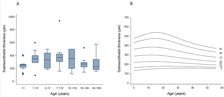
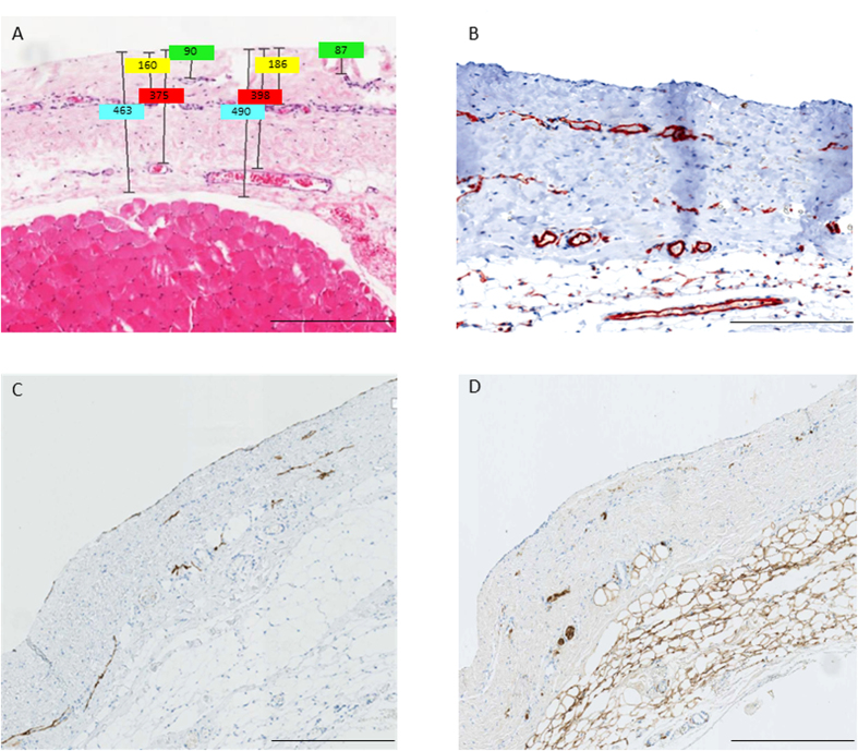
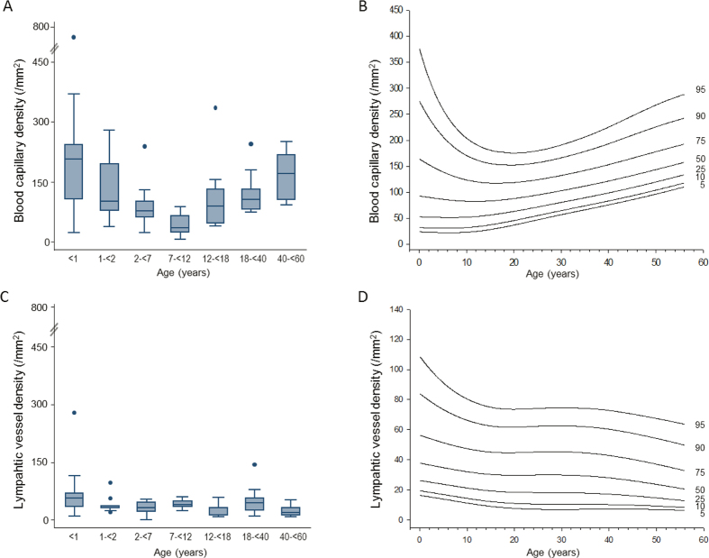
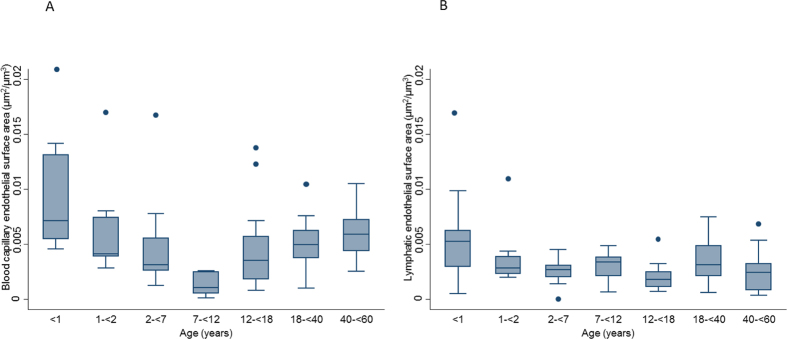
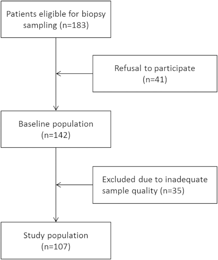
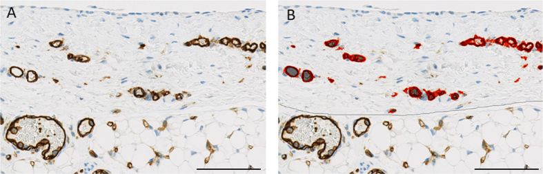
The peritoneum plays an essential role in preventing abdominal frictions and adhesions and can be utilized as a dialysis membrane. Its physiological ultrastructure, however, has not yet been studied systematically. 106 standardized peritoneal and 69 omental specimens were obtained from 107 patients (0.1-60 years) undergoing surgery for disease not affecting the peritoneum for automated quantitative histomorphometry and immunohistochemistry. The mesothelial cell layer morphology and protein expression pattern is similar across all age groups. Infants below one year have a thinner submesothelium; inflammation, profibrotic activity and mesothelial cell translocation is largely absent in all age groups. Peritoneal blood capillaries, lymphatics and nerve fibers locate in three distinct submesothelial layers. Blood vessel density and endothelial surface area follow a U-shaped curve with highest values in infants below one year and lowest values in children aged 7-12 years. Lymphatic vessel density is much lower, and again highest in infants. Omental blood capillary density correlates with parietal peritoneal findings, whereas only few lymphatic vessels are present. The healthy peritoneum exhibits major thus far unknown particularities, pertaining to functionally relevant structures, and subject to substantial changes with age. The reference ranges established here provide a framework for future histomorphometric analyses and peritoneal transport modeling approaches.
Conflict of interest statement
The authors declare no competing financial interests.
Figures
References
-
- Di Paolo N. & Sacchi G. Atlas of peritoneal histology. Perit Dial Int. 20 Suppl 3, S5–96 (2000). - PubMed
-
- Tenckhoff H. & Schechter H. A bacteriologically safe peritoneal access device. Trans Am Soc Artif Intern Organs. 14, 181–187 (1968). - PubMed
-
- Nolph K. D. et al.. Continuous ambulatory peritoneal dialysis: three-year experience at one center. Ann Intern Med. 92, 609–613 (1980). - PubMed
Publication types
MeSH terms
Substances
LinkOut - more resources
Full Text Sources
Other Literature Sources
