

Regulation of epithelial-mesenchymal transition through epigenetic and post-translational modifications
- PMID: 26905733
- PMCID: PMC4765192
- DOI: 10.1186/s12943-016-0502-x
Regulation of epithelial-mesenchymal transition through epigenetic and post-translational modifications
Abstract
The epithelial to mesenchymal transition (EMT) is a biological process in which a non-motile epithelial cell changes to a mesenchymal phenotype with invasive capacities. This phenomenon has been well documented in multiple biological processes including embryogenesis, fibrosis, tumor progression and metastasis. The hallmark of EMT is the loss of epithelial surface markers, most notably E-cadherin, and the acquisition of mesenchymal markers including vimentin and N-cadherin. The downregulation of E-cadherin during EMT can be mediated by its transcriptional repression through the binding of EMT transcription factors (EMT-TFs) such as SNAIL, SLUG and TWIST to E-boxes present in the E-cadherin promoter. Additionally, EMT-TFs can also cooperate with several enzymes to repress the expression of E-cadherin and regulate EMT at the epigenetic and post- translational level. In this review, we will focus on epigenetic and post- translational modifications that are important in EMT. In addition, we will provide an overview of the various therapeutic approaches currently being investigated to undermine EMT and hence, the metastatic progression of cancer as well.
Figures


 indicates inhibition. Purple arrowhead indicates translocation.)
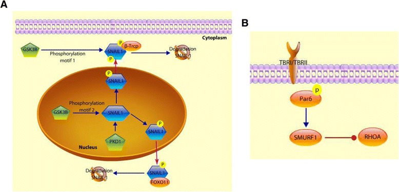
 indicates inhibition. Purple arrowhead indicates translocation.) a. SNAIL phosphorylation that suppresses EMT. GSK-3β phosphorylates SNAIL at two consecutive motifs. First, the phosphorylation at the second motif induces the cytoplasmic translocation of SNAIL from the nucleus. In the cytoplasm, SNAIL is phosphorylated on motif 1, and this phosphorylation is recognized by β-Trcp which targets it for proteosomal degradation of SNAIL. PKD1 is another kinase that phosphorylates SNAIL so it can be recognized by β-Trcp and FOXO11 that target it for proteosomal degradation. b. Mechanisms of EMT activation mediated by TGFβR. The activation of TGFβR results in the phosphorylation of Par6, and in turn activation of SMURF1 that targets RHOA degradation by the proteasome, which contributes to the disassembly of the tight junctions
 indicates inhibition.) a. Sumoylation of FOXM1 represses the expression of miR-200b/c, which normally acts as a tumor suppressor and reduces the expression of transcription factors ZEB1 and ZEB2.As a consequence epithelial phenotype is inhibited. b. Sumoylation of SIP1 decreases its transcription, which in turn prevents recruitment of CtBP to E-cadherin promoter. As a consequence, CtBP cannot prevent E-cadherin expression, which results in the maintenance of the expression of epithelial genes
References
Publication types
MeSH terms
Substances
LinkOut - more resources
Full Text Sources
Other Literature Sources
Research Materials
