. 2016 Apr 1;129(7):1416-28.
doi: 10.1242/jcs.183426. Epub 2016 Feb 18.

Temporal control of bidirectional lipid-droplet motion in Drosophila depends on the ratio of kinesin-1 and its co-factor Halo

Affiliations

Temporal control of bidirectional lipid-droplet motion in Drosophila depends on the ratio of kinesin-1 and its co-factor Halo

Gurpreet K Arora et al. J Cell Sci. .

Abstract

During bidirectional transport, individual cargoes move continuously back and forth along microtubule tracks, yet the cargo population overall displays directed net transport. How such transport is controlled temporally is not well understood. We analyzed this issue for bidirectionally moving lipid droplets in Drosophila embryos, a system in which net transport direction is developmentally controlled. By quantifying how the droplet distribution changes as embryos develop, we characterize temporal transitions in net droplet transport and identify the crucial contribution of the previously identified, but poorly characterized, transacting regulator Halo. In particular, we find that Halo is transiently expressed; rising and falling Halo levels control the switches in global distribution. Rising Halo levels have to pass a threshold before net plus-end transport is initiated. This threshold level depends on the amount of the motor kinesin-1: the more kinesin-1 is present, the more Halo is needed before net plus-end transport commences. Because Halo and kinesin-1 are present in common protein complexes, we propose that Halo acts as a rate-limiting co-factor of kinesin-1.

Keywords: Bidirectional transport; Drosophila embryos; Kinesin-1; Lipid droplets.


Conflict of interest statement

Competing interests

The authors declare no competing or financial interests.

Figures

Fig. 1.
Fig. 1.
Halo locus promotes net plus-end transport in phase II. (A–E) Lipid-droplet (LD) distribution in embryos, revealed by staining for Jabba (green) and nuclei (blue). (A,B) In wild-type embryos, droplets are broadly distributed in the peripheral cytoplasm in phase I, and move inward in phase IIa. This is followed by outward redistribution during phase IIb and phase III. Panels depict either the entire embryo (A) or a section of the periphery (B); the bar on the right in B indicates the approximate location, orientation and polarity of microtubules in these micrographs. (C) Lipid droplets are mislocalized during phase IIa in ΔhaloAJ embryos (also referred to as halo or 0× halo+ in the rest of the paper). (D,E) Inward droplet accumulation during phase IIa is restored if halo embryos express genomic halo+ transgenes. (F) In situ hybridization for halo mRNA. (G) Anti-HA immunoblotting of wild-type and p[haloHA] embryos, and of pellets from bacteria expressing two different sizes of Halo–HA, based on the two annotated start sites. The high-molecular-mass bands present in both wild-type and p[haloHA] embryos are due to non-specific cross-reactivity and serve as loading controls. Scale bars: 7.5 µm (B–E); 75 µm (A,F). Representative examples of at least three independently performed experiments are shown.
Fig. 2.
Fig. 2.
The onset and degradation of Halo promote transitions between phases. (A) Detection of HA (α-HA) in wild-type (phase IIa, WT) and p[haloHA] embryos. Halo is absent in cleavage-stage embryos (a) and is first detected during late phase I (cycle 13, b), with levels rising into phase IIa (c) and declining in phase IIb (d), and levels are drastically reduced in phase III (e). (B) Immunoblotting for HA by using equal numbers of fixed embryos (same ages as in A) reveals the same transient (less than 1.5 h) pattern. (C) In situ hybridization for halo mRNA. In phase III, halo message is barely detectable in wild-type embryos, but remains high in wech embryos. (D) Immunostaining for HA in phase-III embryos reveals increased Halo levels in wech versus wild-type embryos. (E–G) Staining for Jabba to assess droplet (LD) distribution in phase-III embryos. (E) Lipid droplets are present throughout the embryo periphery in the wild type. (Fa) In embryos lacking both the maternal and zygotic supply of wech, droplets remain inward, as in phase IIa. (Fb) In embryos lacking only the maternal supply of wech, the droplet distribution resembles that of wild type. (G) In embryos lacking both wech and halo, droplets are peripheral. (H) Wild-type embryos in early phase III that had been injected during late phase IIa. Arrows indicate injection site. (a,b) Embryos that had been injected with halo mRNA remain transparent around the injection site. Buffer-injected embryos have an opaque periphery everywhere. Scale bars: 75 µm. Representative examples of at least three independently performed experiments are shown.
Fig. 3.
Fig. 3.
Halo levels are crucial for the temporal regulation of droplet transport. (A,D) Staining of Jabba (green) in embryos classified into eight temporal classes (t0 to t7) – cycle 12 (t0, phase I), cycle 13 (t1, phase-I to phase-IIa transition), cycle 14 (t2–t6, phase IIa), late cycle 14 (t7, phase-IIa to phase-IIb transition). Nuclei are blue. (A) In wild-type (WT) embryos, the droplet population continuously shifts inwards from t1 onwards, followed by a reversal at t7. (B,C) Droplet distribution in wild-type embryos at t1 and t6. The fraction of the droplet population (B) and the cumulative population of the droplets (C) along the apical–basal axis of embryos as a function of distance from the apical edge of the nuclei. (D) In halo embryos, the droplet population moves outwards over time. (E) Average droplet position as a function of time for wild-type and halo embryos. The apparent inward shift for halo embryos during t6 and t7 is likely to result from droplets being excluded by increasingly larger nuclei. (B,E) Data are presented as mean±s.d. of three embryos. At each age class, apart from t0, there is a significant difference in the average droplet position between wild-type and halo embryos (Table S1). In the wild type, average droplet position is significantly different between t3 and t5 (P≤0.001, see Table S1), as assessed by t-test. Plots in B,C,E are based on the same dataset. (F) Difference in the average position of the droplets between wild-type and halo embryos for each age class. (G) Immunoblotting for HA by using equal numbers of haloHA embryos from t1 to t7. Wild-type embryos serve as negative control. Scale bars: 7.5 µm (A,D). Representative examples of at least three independently performed experiments are shown with the exception of the experiment shown in G, which was done twice.
Fig. 4.
Fig. 4.
Halo levels need to surpass a threshold to affect droplet transport. (A,B) Droplet (LD) distribution in embryos expressing 1× halo+ and 4× halo+, as revealed by staining for Jabba (green). Nuclei are blue. (C) Average droplet position over time in embryos of different halo+ dosages. The data for 0× and 2× halo+ embryos are the same as in Fig. 3E. There is no statistical significant difference between all genotypes at t0 (Table S1). At t1, the droplets move inwards earlier in embryos expressing 4× halo+, whereas in embryos expressing 1× halo+, the droplet position is indistinguishable from 0× halo+ embryos (Table S1). (D) In situ hybridization for halo message in embryos expressing 1×, 2× or 4× halo+. (E) Immunoblot for HA by using phase-IIa embryos expressing 1× or 2× haloHA in a wild-type or halo background. Halo protein levels change with gene dosage. (F) Staining for Jabba in t1 and t4 embryos expressing 0×, 1× or 2× haloHA in a halo background. (G) At t1, the average droplet position is indistinguishable for 0×, 1× or 2× haloHA embryos. At t4, 0× and 1× haloHA embryos remain indistinguishable, whereas 2× haloHA embryos have diverged, displaying an inward shift (see Table S2 for P-values). (C,G) Data are presented as mean±s.d. of three embryos. Scale bars: 7.5 µm (A,B,F); 75 µm (D). Representative examples of at least three independently performed experiments are shown.
Fig. 5.
Fig. 5.
Halo does not control transport by regulating transcription or acting solely through LSD-2. (A) Staining for HA in wild-type and haloHA embryos. Halo localizes to both nuclei and cytoplasm. (B) Staining for Jabba (red) and HA (green) in centrifuged, heat-fixed wild-type and haloHA embryos. Some Halo associates with the droplet layer (marked by Jabba). Wild-type embryos act as negative control. (C) Equal amounts of protein from haloHA lipid-droplet samples (LD) and whole-embryo lysate (EL) analyzed by immunoblotting. The droplet fraction is highly enriched for the droplet protein LSD-2 and depleted for the cytoplasmic protein tubulin. Because the droplet proteome is less complex than that of the lysate, the increased Halo signal in the LD lane indicates that Halo is relatively more abundant in the droplet fraction than in the embryo as a whole. As the enrichment is much less than that for LSD-2 (at these exposures, LSD-2 signal in the lysate is undetectable), Halo must also be present elsewhere. (D) Injection of wild-type (a) embryos with α-amanitin followed by re-injection with halo mRNA (c) or buffer alone (b). α-Amanitin causes droplets to move outwards (b), similar to the situation in halo embryos (opaque periphery), and on re-injection with halo mRNA, the droplets move inward near the injection site (transparent region), similar to what is seen in panel Da. Arrow indicates injection site. (E) Staining of Jabba in LSD-2 and LSD-2;halo embryos. In halo;LSD-2 double mutants, droplets are shifted outward, compared to their position in LSD-2 embryos. Scale bars: 7.5 µm (E, bottom row); 75 µm (A,B,D,E, top row). Representative examples of at least three independently performed experiments are shown.
Fig. 6.
Fig. 6.
The kinesin-1:Halo ratio is crucial for correct droplet transport. (Aa) The Khc–GFP signal reveals the broad cytoplasmic distribution of kinesin-1. Wild-type embryos serve as negative control. (Ab,B) Staining of kinesin-1 in uncentrifuged (Ab) and centrifuged wild-type embryos (B). Kinesin is broadly distributed in the cytoplasm (A), and a small fraction associates with the droplet layer (B). (C,D) Staining of Jabba in embryos expressing different ratios of kinesin-1 to Halo, age classes t1 and t5. (C) Global droplet distribution in 1× Khc; 1× halo+ embryos is similar to that of wild-type (2× Khc; 2× halo+) embryos at both t1 and t5 (P-value was not significant, Table S3). (D) Kinesin-1 overexpression attenuates net motion in the plus-end direction. The droplet population shifts inward as additional copies of halo are added. (E,F) Average droplet position in the genotypes described in C,D. (G) Staining for Jabba reveals the droplet distribution in 1× Khc; 0× halo+ embryos. The distribution is similar to that observed in halo embryos. (H) Average position of droplets in 1× Khc; 0× halo+ and 2× Khc; 0× halo+ embryos. At each time point, the droplet position is indistinguishable (P-value was not significant, calculated by performing a t-test between the two genotypes at each age class). (E,F,H) Data are presented as mean±s.d. of three embryos. Scale bars: 75 µm (A,B); 7.5 µm (C,D,G). Representative examples of at least three independently performed experiments are shown.
Fig. 7.
Fig. 7.
Kinesin-1 sets the threshold for Halo. (A) Staining for Jabba in 2× Khc; 1× haloHA (repeated from Fig. 4F for side-by-side comparison) or 1× Khc; 1× haloHA embryos, with 1× haloHA being the only source of Halo function. (B) Average droplet position for the genotypes shown in A. At t1, the average droplet position in both genotypes is similar (P-value was not significant, Table S5). At t4, droplets had shifted inward for 1× Khc embryos but outward for 2× Khc embryos. Data are presented as mean±s.d. of three embryos. (C) Immunoblotting for Khc in embryos at t1, t4, t7 and phase III. Kinesin-1 levels are similar in wild-type (WT) and halo embryos. Tubulin serves as loading control. (D,E) HA detection by immunoblotting and immunostaining in embryos expressing 2×, 1× and ∼8× Khc. (D) Halo distribution is similar in all genotypes. (E) Blotting for Khc shows the kinesin-1 levels. α-Tubulin is the loading control. Halo levels do not change with Khc dosage. (F) Immunoprecipitation of HA using lysates from wild-type and haloHA embryos. Kinesin-1 co-purifies with Halo. (G) Immunoprecipitation of Khc using lysates from wild-type and haloHA embryos. Halo co-purifies with kinesin-1. Scale bars: 7.5 µm (A); 75 µm (D). Representative examples of at least three independently performed experiments are shown.
Fig. 8.
Fig. 8.
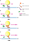
Model of temporal control of droplet transport. During phase IIa, plus-end run lengths are not upregulated in the absence of Halo (A) or when only a subset of kinesins are activated by Halo binding (B). This lack of upregulation results in net transport in the minus-end direction. (C) Once Halo levels surpass the threshold to bind all kinesins and activate them, plus-end travel distances lengthen, resulting in net plus-end transport. (D) Fewer kinesins per droplet allow full activation at lower Halo levels. A reduction in kinesin-1 levels also reduces the number of active dynein motors (Shubeita et al., 2008). This model does not attempt to incorporate added complexities of how Halo might affect minus-end run lengths (Gross et al., 2003).

References

    1. Aspengren S., Hedberg D., Sköld H. N. and Wallin M. (2009). New insights into melanosome transport in vertebrate pigment cells. Int. Rev. Cell Mol. Biol. 272, 245-302. 10.1016/S1937-6448(08)01606-7 - DOI - PubMed
    1. Block S. M. (2007). Kinesin motor mechanics: binding, stepping, tracking, gating, and limping. Biophys. J. 92, 2986-2995. 10.1529/biophysj.106.100677 - DOI - PMC - PubMed
    1. Bonneaud N., Savare J., Berta P. and Girard F. (2003). SNCF, a SoxNeuro interacting protein, defines a novel protein family in Drosophila melanogaster. Gene 319, 33-41. 10.1016/S0378-1119(03)00795-9 - DOI - PubMed
    1. Brendza K. M., Rose D. J., Gilbert S. P. and Saxton W. M. (1999). Lethal kinesin mutations reveal amino acids important for ATPase activation and structural coupling. J. Biol. Chem. 274, 31506-31514. 10.1074/jbc.274.44.31506 - DOI - PMC - PubMed
    1. Brendza R. P., Sheehan K. B., Turner F. R. and Saxton W. M. (2000). Clonal tests of conventional kinesin function during cell proliferation and differentiation. Mol. Biol. Cell 11, 1329-1343. 10.1091/mbc.11.4.1329 - DOI - PMC - PubMed

Publication types

MeSH terms

LinkOut - more resources