Review
. 2016 May-Jun;51(3):162-84.
doi: 10.3109/10409238.2016.1143913. Epub 2016 Feb 24.

PP2A as a master regulator of the cell cycle

Affiliations
Review

PP2A as a master regulator of the cell cycle

Nathan Wlodarchak et al. Crit Rev Biochem Mol Biol. 2016 May-Jun.

Abstract

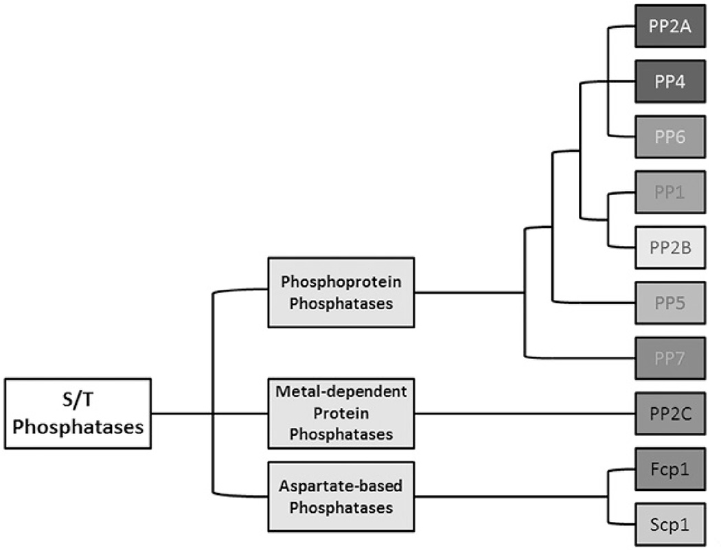
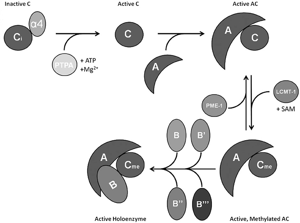
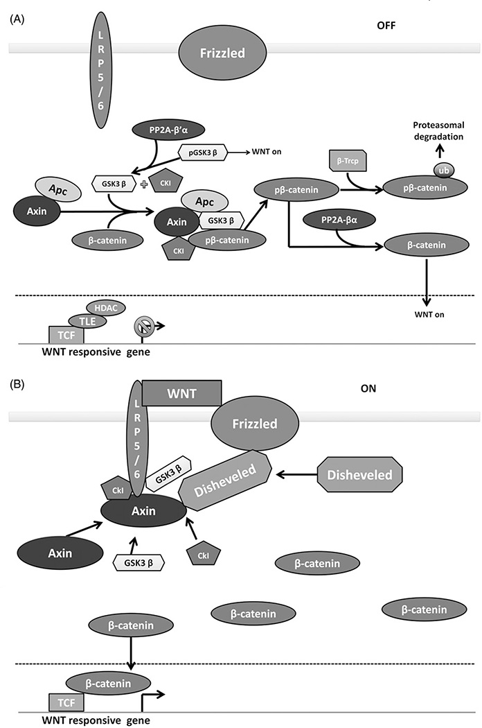
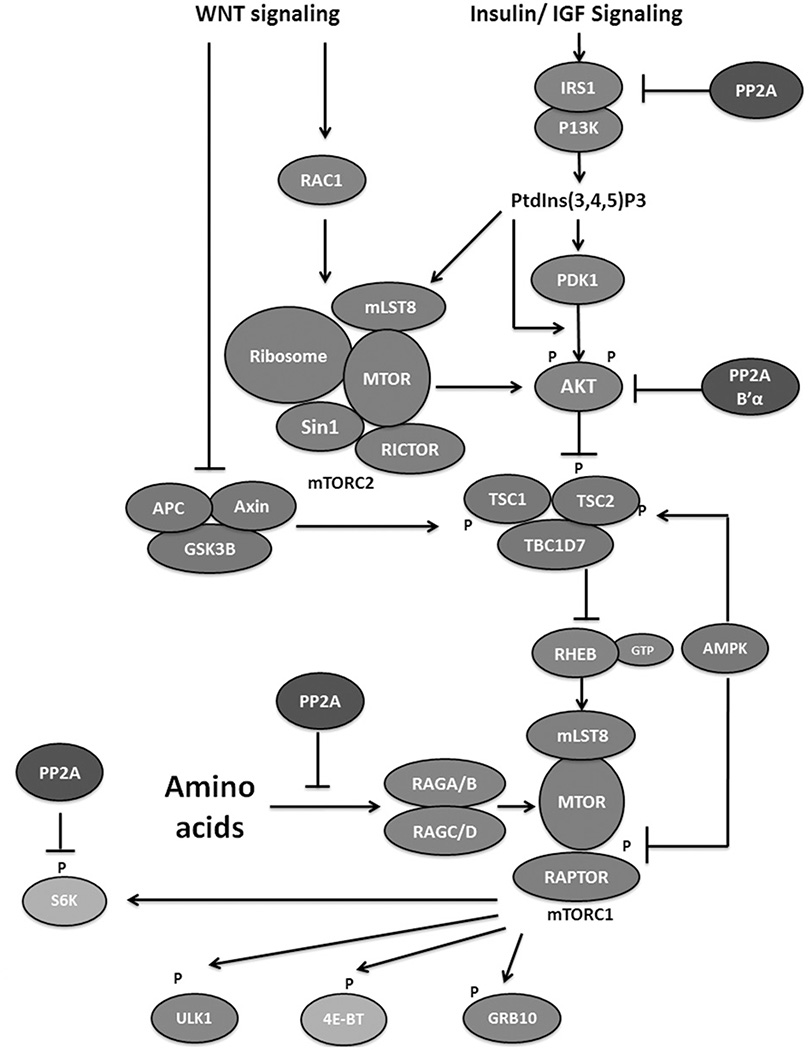
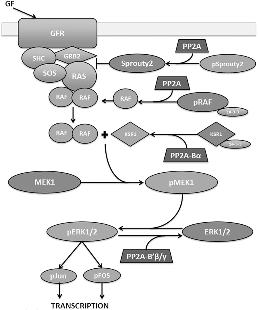
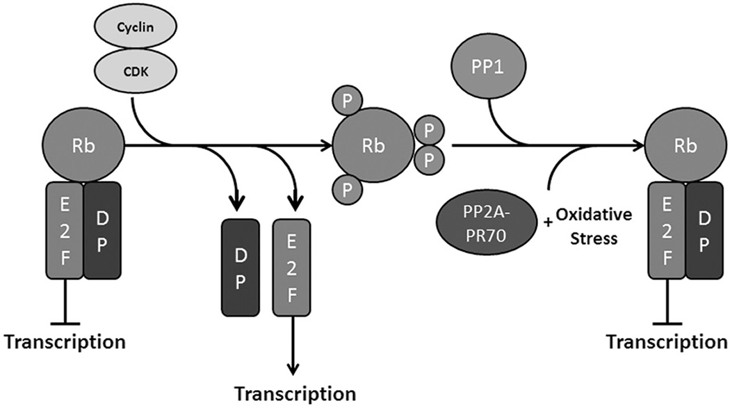
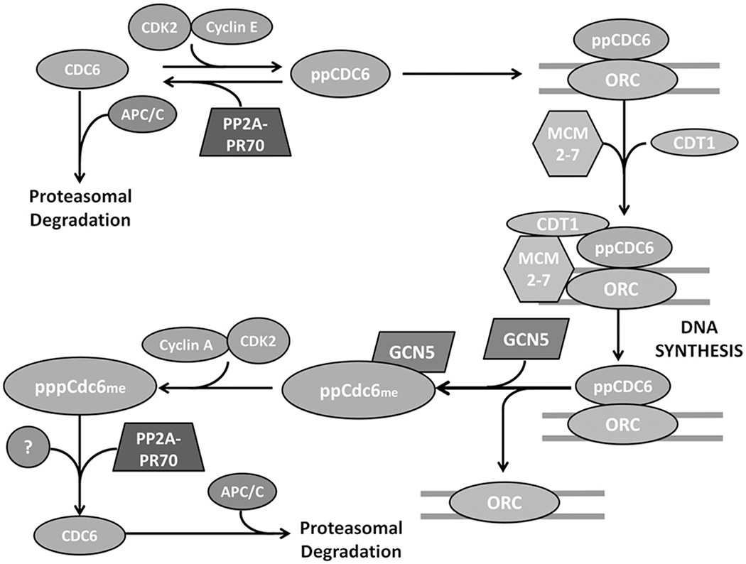
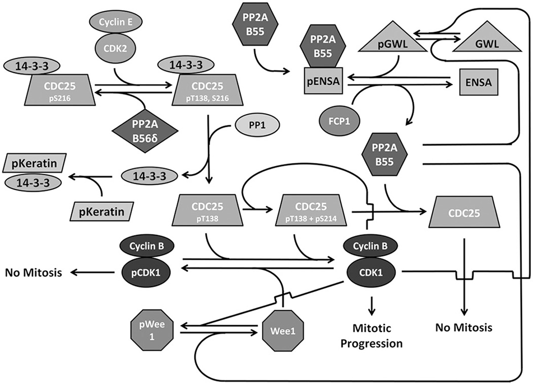
Protein phosphatase 2A (PP2A) plays a critical multi-faceted role in the regulation of the cell cycle. It is known to dephosphorylate over 300 substrates involved in the cell cycle, regulating almost all major pathways and cell cycle checkpoints. PP2A is involved in such diverse processes by the formation of structurally distinct families of holoenzymes, which are regulated spatially and temporally by specific regulators. Here, we review the involvement of PP2A in the regulation of three cell signaling pathways: wnt, mTOR and MAP kinase, as well as the G1→S transition, DNA synthesis and mitotic initiation. These processes are all crucial for proper cell survival and proliferation and are often deregulated in cancer and other diseases.

Keywords: Cancer; PP2A; cell cycle; cell division; mitosis; phosphatase.


Figures

Figure 1
Figure 1
Serine/threonine phosphatases are classified based on biochemical mechanism. They are divided into three families, the aspartate-based phosphatases, the metal-dependent protein phosphatases and the phosphoprotein phosphatases. The phosphoprotein phosphatases have similar active site configurations and require catalytic metal ions in the active site. PP2A is a member of this family. Adapted from Stanevich (2013). (see colour version of this figure at www.informahealthcare.com/bmg).
Figure 2
Figure 2
PP2A biogenesis and holoenzyme assembly is regulated by unique factors. α4 protects inactive PP2Ac from ubiquitination by MID1. Activating metal ions are loaded by PTPA, and active PP2Ac binds to the scaffold subunit (A). The C-terminal tail of PP2Ac can be methylated by LCMT-1 and reversed by PME-1. Active, methylated PP2A-AC can then form holoenzymes with one B subunit. These available B subunits are divided into four families: B, B′, B″ and B‴, each with unique characteristics and regulation. (see colour version of this figure at www.informahealthcare.com/bmg).
Figure 3
Figure 3
PP2A positively and negatively regulates the Wnt signaling pathway. (A) Wnt OFF. In the absence of Wnt signaling, a complex of Axin, Apc, GSK3β and CK1 phosphorylate β-catenin, targeting it for proteasomal degradation. PP2A-B′α promotes β-catenin degradation by removing an inhibitory phosphorylation on GSK3β. PP2A-Bα can directly dephosphorylate β-catenin, promoting the activation of wnt responsive genes. (B) Wnt signaling ON. In the presence of Wnt ligand, Wnt receptors LRP5/6 and frizzled sequester the Axin, GSK3β and CK1, preventing the phosphorylation of β-catenin. β-Catenin accumulates and translocates to the nucleus, promoting the transcription of Wnt responsive genes. Figure adapted from MacDonald et al. (2009). (see colour version of this figure at www.informahealthcare.com/bmg).
Figure 4
Figure 4
PP2A negatively regulates the mTOR signaling pathway. The mTOR complexes are colored blue. Proteins involved in mTOR inhibition and activation are colored red and green, respectively with PP2A in purple. Downstream factors inhibiting and stimulating growth are colored magenta and teal, respectively. Growth factors stimulate the mTOR pathway via inhibiting the function of the TSC complex that inhibits mTOR activation. Wnt signaling can inhibit the TSC complex or directly stimulate mTORC2. Amino acids can also stimulate mTOR activity. PP2A inhibits the mTOR pathway by inhibiting IRS1 in the insulin signaling pathway or MAP4K3 in the amino acid pathway, or by inhibiting AKT function. PP2A can also reverse mTOR phosphorylation of S6K. Figure adapted from Shimobayshi & Hall (2014). (see colour version of this figure at www.informahealthcare.com/bmg).
Figure 5
Figure 5
PP2A positively and negatively regulates the MAPK signaling pathway. Growth factors stimulate a complex of proteins: SHC, GRB2 and SOS to assemble on a growth factor receptor. This complex activates Ras which starts a signal cascade from activation of Raf, to activation of MEK, ERK and eventually the transcription factors that activate the transcription of growth related genes. PP2A can activate Raf by dephosphorylating S259 and causing 14–3-3 release. PP2A-Bα dephosphorylates S392 of KSR1, which leads to dissociation of 14–3-3 from KSR1, essential for MEK1 activation. PP2A negatively regulates MAPK upstream by activating Sprouty2, which inhibits GRB2 and subsequent RAS complex formation. PP2A-B′β/B′γ can directly dephosphorylate ERK1/2 downstream of the signaling cascade, thereby inactivating it. Factors promoting cell division are shown in green and those opposing cell division are shown in red. Figure adapted from McCubrey et al. (2007). (see colour version of this figure at www.informahealthcare.com/bmg).
Figure 6
Figure 6
Rb phosphorylation promotes transcription of E2F responsive genes. Rb normally binds E2F transcription factors and their dimerization partners. When phosphorylated by cyclin/CDK heterodimers, Rb loses affinity for E2F and free E2F is allowed to promote transcription. Rb is dephosphorylated at the end of mitosis, allowing reassociation with E2F. Normally, PP1 dephosphorylates Rb at the end of mitosis, but PP2A-PR70 can dephosphorylate Rb under oxidative stress conditions. (see colour version of this figure at www.informahealthcare.com/bmg).
Figure 7
Figure 7
Cdc6 is necessary for assembly of the pre-replication complex and subsequent DNA synthesis. In G0, Cdc6 is ubiquinated by the anaphase promoting complex/cyclosome (APC/C) and degraded by the proteasome. In G1, Cyclin E/CDK2 phosphorylates Cdc6 on S54 and S74, protecting it from degradation. Cdc6 is translocated into the nucleus where it binds the origin recognition complex and is required to recruit Cdt1 and MCM2–7 and form the prereplication complex. After firing of the origins, Cdc6 is methylated by GCN5 causing its dissociation from the ORC. Cdc6 is then phosphorylated on S106 by Cyclin A/CDK2 and translocated to the cytoplasm. PP2A-PR70 is thought to dephosphorylate Cdc6 either at this point in G2 and/or in G1, ensuring Cdc6 destruction and regulating DNA synthesis. Figure adapted from Mumby (2009). (see colour version of this figure at www.informahealthcare.com/bmg).
Figure 8
Figure 8
PP2A negatively regulates the cell cycle through CDC25 and Wee1. In G2, Greatwall, Fcp and Wee1 are dephosphorylated, keeping PP2A-B55 active and CDK1 inactive. CDC25 is phosphorylated at S216, allowing 14–3-3 association, holding it inactive. At the transition from G2 to M, CDC25 is phosphorylated at T138, weakening 14–3-3 binding and allowing dissociation with subsequent binding to a phospho-keratin pool. The now exposed S216 can be dephosphorylated by PP1 activating CDC25. Active CDC25 dephosphorylates CDK1 at T14 and Y15 thereby activating it. The active CDK1 can then phosphorylate several substrates required for mitotic progression. In addition, CDK1 participates in several positive feedback loops. It phosphorylates Wee1, preventing direct inactivation, and it phosphorylates CDC25 at S214, increasing its affinity for PP1 and allowing for more CDC25 activation. Furthermore, CDK1 can prevent CDC25 inactivation by PP2A-B55 by phosphorylating Greatwall, which in turn phosphorylates ENSA, which binds to PP2A-B55 and keeps it inactive. CDK1 also phosphorylates FCP1, preventing it from dephosphorylating ENSA and releasing PP2A-B55. Figure adapted from Johnson & Kornbluth (2012) and Hegarat et al. (2014). (see colour version of this figure at www.informahealthcare.com/bmg).

References

    1. Abdelmohsen K, Srikantan S, Yang X, et al. Ubiquitin-mediated proteolysis of HuR by heat shock. EMBO J. 2009;28:1271–1282. - PMC - PubMed
    1. Adams DG, Coffee RL, Jr, Zhang H, et al. Positive regulation of Raf1-MEK1/2-ERK1/2 signaling by protein serine/threonine phosphatase 2A holoenzymes. J Biol Chem. 2005;280:42644–42654. - PubMed
    1. Adler HT, Nallaseth FS, Walter G, Tkachuk DC. HRX leukemic fusion proteins form a heterocomplex with the leukemia-associated protein SET and protein phosphatase 2A. J Biol Chem. 1997;272:28407–28414. - PubMed
    1. Afonso O, Matos I, Pereira AJ, et al. Feedback control of chromosome separation by a midzone Aurora B gradient. Science. 2014;345:332–336. - PMC - PubMed
    1. Ahn JH, Mcavoy T, Rakhilin SV, et al. Protein kinase A activates protein phosphatase 2A by phosphorylation of the B56delta subunit. Proc Natl Acad Sci USA. 2007a;104:2979–2984. - PMC - PubMed

Substances

LinkOut - more resources