Review
. 2016;11(5):513-30.
doi: 10.2217/nnm.16.3. Epub 2016 Feb 23.

Nanotechnology in diagnosis and treatment of coronary artery disease

Affiliations
Review

Nanotechnology in diagnosis and treatment of coronary artery disease

Mahdi Karimi et al. Nanomedicine (Lond). 2016.

Abstract

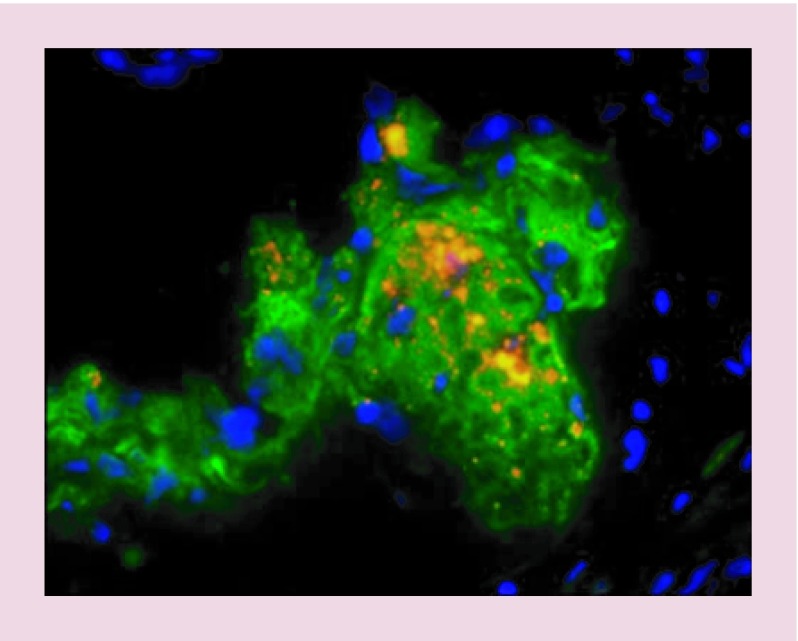
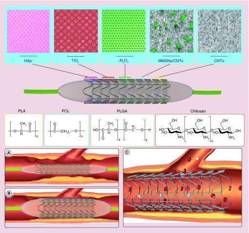
Nanotechnology could provide a new complementary approach to treat coronary artery disease (CAD) which is now one of the biggest killers in the Western world. The course of events, which leads to atherosclerosis and CAD, involves many biological factors and cellular disease processes which may be mitigated by therapeutic methods enhanced by nanotechnology. Nanoparticles can provide a variety of delivery systems for cargoes such as drugs and genes that can address many problems within the arteries. In order to improve the performance of current stents, nanotechnology provides different nanomaterial coatings, in addition to controlled-release nanocarriers, to prevent in-stent restenosis. Nanotechnology can increase the efficiency of drugs, improve local and systematic delivery to atherosclerotic plaques and reduce the inflammatory or angiogenic response after intravascular intervention. Nanocarriers have potential for delivery of imaging and diagnostic agents to precisely targeted destinations. This review paper will cover the current applications and future outlook of nanotechnology, as well as the main diagnostic methods, in the treatment of CAD.

Keywords: atherosclerosis; coronary artery disease; nanocarriers; nanotechnology; restenosis; stent coatings; vulnerable plaque.


Conflict of interest statement

Financial & competing interests disclosure MR Hamblin was supported by US NIH Grant R01AI050875. The authors have no other relevant affiliations or financial involvement with any organization or entity with a financial interest in or financial conflict with the subject matter or materials discussed in the manuscript apart from those disclosed. No writing assistance was utilized in the production of this manuscript.

Figures

<b>Figure 1.</b>
Figure 1.. Schematic structure of important nanoparticles.
<b>Figure 2.</b>
Figure 2.. The liposomal nanoparticle with prednisolone phosphate stored in a macrophages of iliofemoral plaques.
(A) First row illustrates the plaque cells cured by LN-PLP marked for cell nuclei (DAPI), macrophages (CD68) and liposome-coating PEG. In the second row, magnified images of isolated cells are shown. (B) Third row shows CD68 cells from a plaque cured by saline, but there is no positivity for PEG. LN-PLP: Liposomal nanoparticle with prednisolone phosphate. Reproduced with permission from [39], © (2015) Nanomedicine: Nanotechnology, Biology and Medicine.
<b>Figure 3.</b>
Figure 3.. Delivery of tetrahydrobiopterin (BH4) by liposome nanocarrier.
(A) Improved stability of BH4 in comparison with unencapsulated BH4 after 24 h; p = 0.017; (B) BH4 concentration in ligated artery increased by liposomal delivery; p = 0.04; (C) superoxide concentration in ligated artery with targeted liposomes decreased; (D) the plaque burden was decreased by BH4 liposomal delivery in the mice ligated left carotid artery fed by 7-day high fat diet (plaque and lumen are marked by red and blue, respectively) scale bars = 100 μm; (E) the area of plaque; p = 0.0015; (F) there is no alteration in lipid metabolism via liposome deliver; p = 0.47 and 0.11, respectively). BH4: Tetrahydrobiopterin; DHE: Dihydroethidium; FBS: Fetal bovine serum; LCA: Left common carotid artery; RCA: Right common carotid artery. Reproduced with permission from [40], © (2015) ACS Nano.
<b>Figure 4.</b>
Figure 4.. Paclitaxel-loaded magnetic nanoparticles applied to coronary stents with a uniform magnetic field.
MNPs with PTX doses of 7.5 and 0.75 μg entered into animal bodies under magnetic versus nonmagnetic conditions. The animals sacrificed and the stented carotid segments were harvested 14 days after surgery. The control group did not receive MNP but stented. Verhoeff–van Gieson-stained section of an artery lumen treated with 7.5 μg PTX under magnetic conditions (A) demonstrated versus ‘no treatment’ control (B) (p < 0.05, Dunn's Test Q statistic = 3.7). Original magnification 100×. Morphometric results as neointima/media ratios (C) pictured as a function of the magnetic field application and PTX dose (n ≥ 6). Data are presented as mean ± standard error. MNP: Magnetic nanoparticle; NP: Nanoparticle; PTX: Paclitaxel. Reprinted with permission from [57], © (2010) Proceedings of the National Academy of Sciences of the United States of America.
<b>Figure 5.</b>
Figure 5.. Using quantum dots to image the monocyte-macrophages in atherosclerosis plaque.
The monocyte-macrophages loaded by cell penetrating quantum dots were injected to mice. Injected cells and macrophage marker CD68 are portrayed as orange and green, respectively. Reproduced with permission from [59], © (2010) Current Atherosclerosis Reports.
<b>Figure 6.</b>
Figure 6.. Stent coating structures and positioning in the coronary artery.
Top: porous structure of various ceramic coatings and chemical structures of polymers that have been applied on stents; (A) plaque accumulation in the lumen of coronary artery and delivery of nonexpanded stent; (B) expanded stent via pressure of balloon catheter; (C) expanded stent remains at the site of plaque allowing good blood flow. CNT: Carbon nanotube; HAp: Hydroxyapatite; MMSN: Magnetic mesoporous silica nanoparticle.

References

    1. Brito L, Amiji M. Nanoparticulate carriers for the treatment of coronary restenosis. Int. J. Nanomedicine. 2007;2:143. - PMC - PubMed
    1. Badimon L, Vilahur G. Thrombosis formation on atherosclerotic lesions and plaque rupture. J. Intern. Med. 2014;276(6):618–632. - PubMed
    1. Cominacini L, Garbin U, Mozzini C, et al. The atherosclerotic plaque vulnerability: focus on the oxidative and endoplasmic reticulum stress in orchestrating the macrophage apoptosis in the formation of the necrotic core. Curr. Med. Chem. 2015;22(13):1565–1572. - PubMed
    1. Naghavi M, Libby P, Falk E, et al. From vulnerable plaque to vulnerable patient a call for new definitions and risk assessment strategies: part I. Circulation. 2003;108(14):1664–1672. - PubMed
    1. Libby P, Ridker PM, Hansson GK. Progress and challenges in translating the biology of atherosclerosis. Nature. 2011;473:317–325. - PubMed

Publication types

MeSH terms

LinkOut - more resources