. 2016 Feb 24:6:21408.
doi: 10.1038/srep21408.

Insights into the mechanism of human papillomavirus E2-induced procaspase-8 activation and cell death

Affiliations

Insights into the mechanism of human papillomavirus E2-induced procaspase-8 activation and cell death

Nitu Singh et al. Sci Rep. .

Abstract

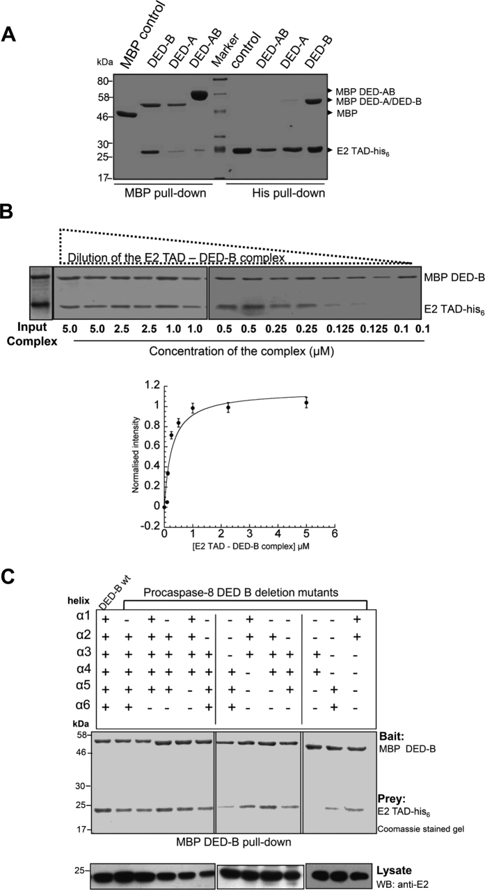
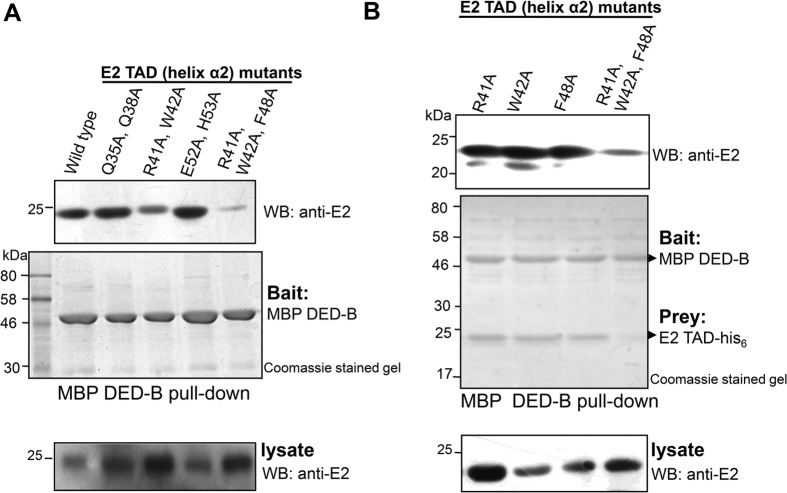
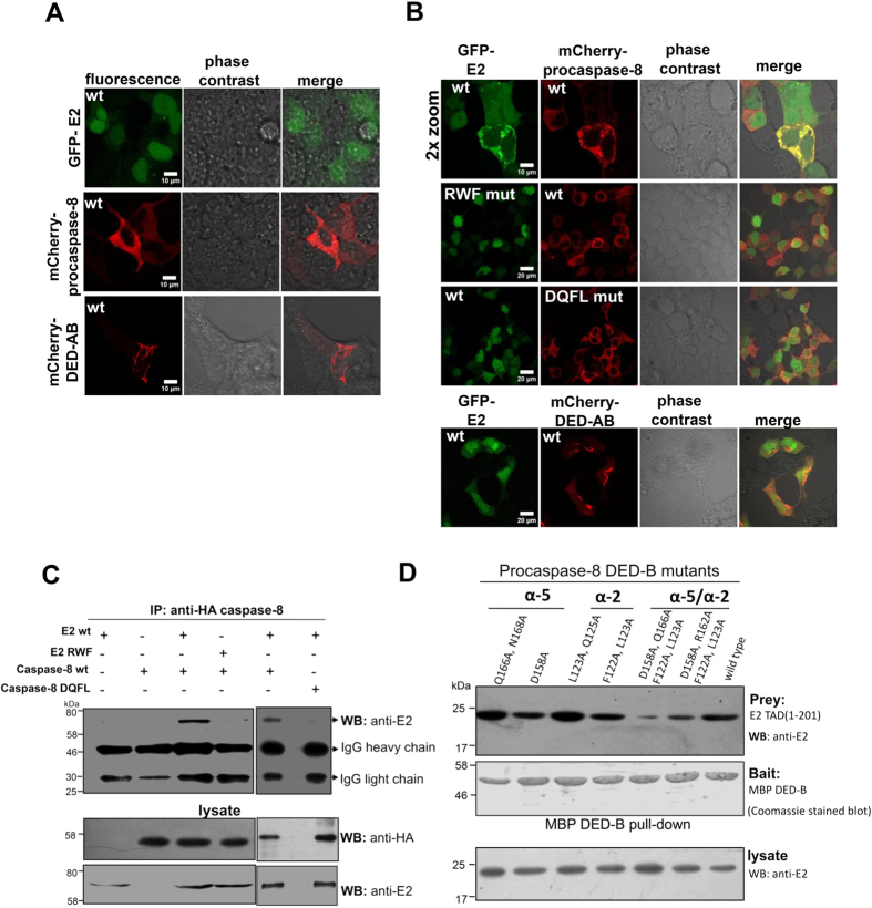
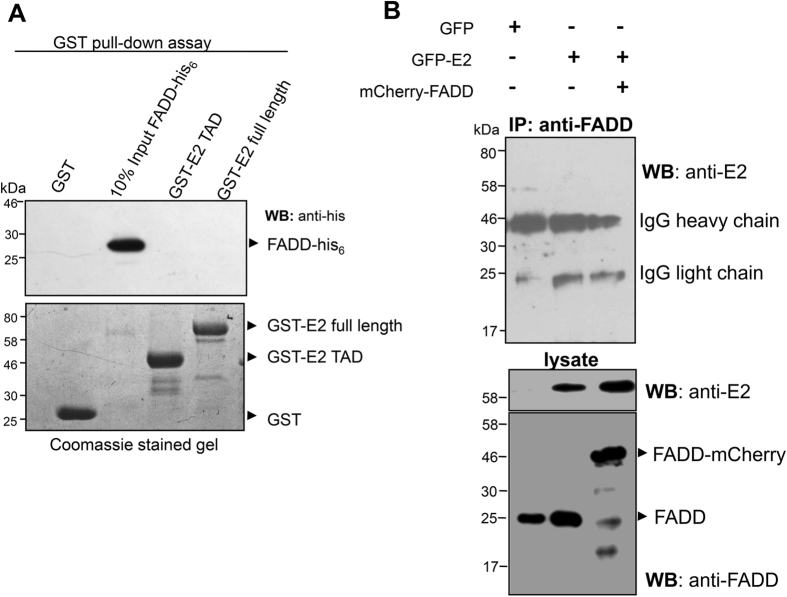
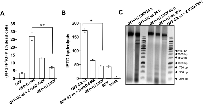
High-risk human papillomavirus (HR-HPV) E2 protein, the master regulator of viral life cycle, induces apoptosis of host cell that is independent of its virus-associated regulatory functions. E2 protein of HR-HPV18 has been found to be involved in novel FADD-independent activation of caspase-8, however, the molecular basis of this unique non-death-fold E2-mediated apoptosis is poorly understood. Here, with an interdisciplinary approach that involves in silico, mutational, biochemical and biophysical probes, we dissected and characterized the E2-procasapse-8 binding interface. Our data demonstrate direct non-homotypic interaction of HPV18 E2 transactivation domain (TAD) with α2/α5 helices of procaspase-8 death effector domain-B (DED-B). The observed interaction mimics the homotypic DED-DED complexes, wherein the conserved hydrophobic motif of procaspase-8 DED-B (F122/L123) occupies a groove between α2/α3 helices of E2 TAD. This interaction possibly drives DED oligomerization leading to caspase-8 activation and subsequent cell death. Furthermore, our data establish a model for E2-induced apoptosis in HR-HPV types and provide important clues for designing E2 analogs that might modulate procaspase-8 activation and hence apoptosis.


Figures

Figure 1
Figure 1. Mapping binding region of E2-procaspase-8 interaction.
(A) Pull-down of E2 TAD and procasapse-8 DED domains. Lanes 1–4 represent MBP pull-down using amylose resin while lanes 6–9 is for reverse his6 pull-down with Ni2+-IDA resin. In lane-1, MBP was checked for binding to E2 TAD-his6, while in lanes 2–4, MBP fused DED-B, DED-A and DED-AB acted as baits. In lanes 6–9, E2 TAD-his6 acted as a bait to monitor the binding of MBP, MBP-tagged DED-AB, DED-A and DED-B respectively. The image is of 12% SDS-PAGE Coomassie-stained gel. (B) Dilution experiment with the isolated E2 TAD – DED-B complex. Top panel-The complex diluted to different final concentrations as indicated were analyzed on SDS-PAGE. The lower panel shows a plot derived from quantitative analysis of background corrected band intensity measured as E2 TAD to DED-B ratio for various dilutions of the complex. The measured intensity is represented as normalized intensity for the data obtained from three independent complex preparations and their dilutions. The error bars show the standard error. (C) MBP pull-down assay of deletion constructs with MBP DED-B and E2 TAD-his6 as bait and prey respectively. 20 μg of the total cell lysate was immunoblotted with anti-E2 antibody to confirm the presence of E2 protein in each sample.
Figure 2
Figure 2. Identification of E2 binding surface for procaspase-8.
(A,B) MBP pull-down assay for E2 wild-type and mutants with procaspase-8 DED-B. E2 TAD wild-type or mutants (prey) as indicated were tested for their binding to MBP-tagged DED-B (bait) using amylose affinity purification. 20 μg of the total lysate immunoblotted with anti-E2 confirmed the presence of E2 TAD in all the samples. The varying amount of protein in the total bacterial lysate is most likely due to difference in the expression level of these proteins in the cell. Although lower in case of single mutants, the in vitro isolated complexes for both the wild-type and single mutant proteins were similar, whereas the combined mutations of these residues significantly affected the complex formation.
Figure 3
Figure 3. E2-procaspase-8 interaction and colocalization.
(A,B) Expression and localization of catalytically inactive procaspase-8 full length, prodomain and HPV18 E2 in transfected cells. HEK293 cells were transfected with the indicated expression constructs and live-cell confocal images were acquired post 20–24 h of transfection. The abbreviations RWF and DQFL stand for: R41A/W42A/F48A and D158A/Q166A/ F122A/L123A respectively. Of the ~100 cells visualized, >45% cells expressing both the wild-type proteins showed re-distribution into co-localized cytoplasmic punctate structures, while it was significantly less, approximately 4–5% for cells expressing the mutant protein. Representative images for each of the transfected population are shown along with the scale bar. (C) Immunoprecipitation of E2-procaspase-8 complex. Extracts from transfected HEK293 cells were immunoprecipitated using an anti-HA antibody followed by immunoblotting using anti-E2. To check the presence of the proteins in the cell extract, 20 μg of the lysate was probed with anti-HA and anti-E2 antibodies. (D) MBP pull-down assay for wild-type E2 TAD and procaspase-8 DED-B mutants. Using amylose resin, MBP-tagged procaspase-8 DED mutants (bait) as indicated were tested for their binding to E2 TAD-his6 (prey) by probing with anti-E2. 20 μg of the total lysate was immunoblotted with anti-E2 to test the presence of E2 TAD in all the samples.
Figure 4
Figure 4. E2-FADD interaction analysis.
(A) Upper panel: Western blot analysis of GST pull-down assay of HPV18 E2 full length or TAD proteins (bait) with recombinant FADD-his6 (prey). Lower panel shows the Coomassie-stained gel for the recombinant GST and GST-fusion proteins. (B) Immunoprecipitation of FADD. Extracts from HEK293 cells co-transfected as described were immunoprecipitated using an anti-FADD antibody followed by immunoblotting using anti-E2. 20 μg of the cell lysate was immunoblotted with anti-FADD and anti-E2 antibodies, to confirm the presence of these proteins in the cellular lysate.
Figure 5
Figure 5. E2-induced cell death analyses.
(A) Percentage of GFP-positive HEK293 dead cells expressing the indicated plasmids was quantified by propidium iodide uptake as described in the Methods section. (B) Caspase-8 activity was measured for the crude extracts isolated from the cells transfected with the plasmids as indicated, using fluorogenic Ac-IETD-AFC substrate. The bars and the error bars represent mean and standard deviation respectively. Statistical analysis using Student’s t-test, *p < 0.05 (A) and p < 0.01 (B). (C) Agarose gel electrophoresis of apoptotic DNA fragments. Total DNA extracted from transfected HEK293 cells was loaded on a 2% agarose gel. A typical DNA ladder consisting of multiples of 180 bp fragments was observed in case of wild-type E2 expressing cells while it was significantly reduced in mutant cells (M stands for 1 kilo-bp DNA marker). In each experiment, cells expressing wild-type E2 incubated with pan-caspase inhibitor (Z-VAD-FMK) was kept as experimental control to monitor that inhibition of the caspases affected substrate cleavage and apoptosis.

References

    1. Scheffner M., Huibregtse J. M., Vierstra R. D. & Howley P. M. The HPV-16 E6 and E6-AP complex functions as a ubiquitin-protein ligase in the ubiquitination of p53. Cell 75, 495–505 (1993). - PubMed
    1. Filippova M., Parkhurst L. & Duerksen-Hughes P. J. The human papillomavirus 16 E6 protein binds to Fas-associated death domain and protects cells from Fas-triggered apoptosis. J Biol Chem 279, 25729–25744 (2004). - PubMed
    1. Blachon S. & Demeret C. The regulatory E2 proteins of human genital papillomaviruses are pro-apoptotic. Biochimie 85, 813–819 (2003). - PubMed
    1. Desaintes C., Demeret C., Goyat S., Yaniv M. & Thierry F. Expression of the papillomavirus E2 protein in HeLa cells leads to apoptosis. EMBO J 16, 504–514 (1997). - PMC - PubMed
    1. Blachon S., Bellanger S., Demeret C. & Thierry F. Nucleo-cytoplasmic shuttling of high-risk HPV E2 proteins induces apoptosis. J. Biol. Chem. 280, 36088–36098 (2005). - PubMed

Publication types

LinkOut - more resources