. 2016 Feb 22:16:134.
doi: 10.1186/s12885-016-2185-5.

Transketolase-like 1 ectopic expression is associated with DNA hypomethylation and induces the Warburg effect in melanoma cells

Affiliations

Transketolase-like 1 ectopic expression is associated with DNA hypomethylation and induces the Warburg effect in melanoma cells

Aparna Jayachandran et al. BMC Cancer. .

Abstract

Background: The metabolism of cancer cells is often reprogrammed by dysregulation of metabolic enzymes. Transketolase-like 1 (TKTL1) is a homodimeric transketolase linking the pentose-phosphate pathway with the glycolytic pathway. It is generally silenced at a transcriptional level in somatic tissues. However, in human cancers its expression is associated with the acquisition of a glycolytic phenotype (the Warburg effect) by cancer cells that contributes to the progression of malignant tumors. In melanoma, defective promoter methylation results in the expression of genes and their products that can affect the tumor cell's phenotype including the modification of immune and functional characteristics. The present study evaluates the role of TKTL1 as a mediator of disease progression in melanoma associated with a defective methylation phenotype.

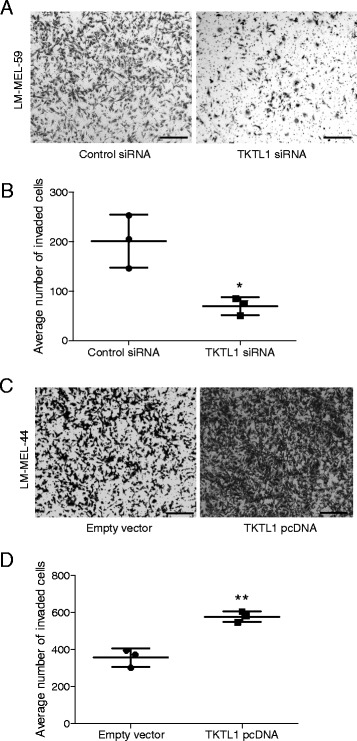
Methods: The expression of TKTL1 in metastatic melanoma tumors and cell lines was analysed by qRT-PCR and immunohistochemistry. The promoter methylation status of TKTL1 in melanoma cells was evaluated by quantitative methylation specific PCR. Using qRT-PCR, the effect of a DNA demethylating agent 5-aza-2'-deoxycytidine (5aza) on the expression of TKTL1 was examined. Biochemical and molecular analyses such as glucose consumption, lactate production, invasion, proliferation and cell cycle progression together with ectopic expression and siRNA mediated knockdown were used to investigate the role of TKTL1 in melanoma cells.

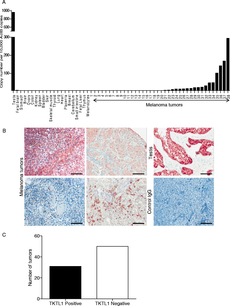
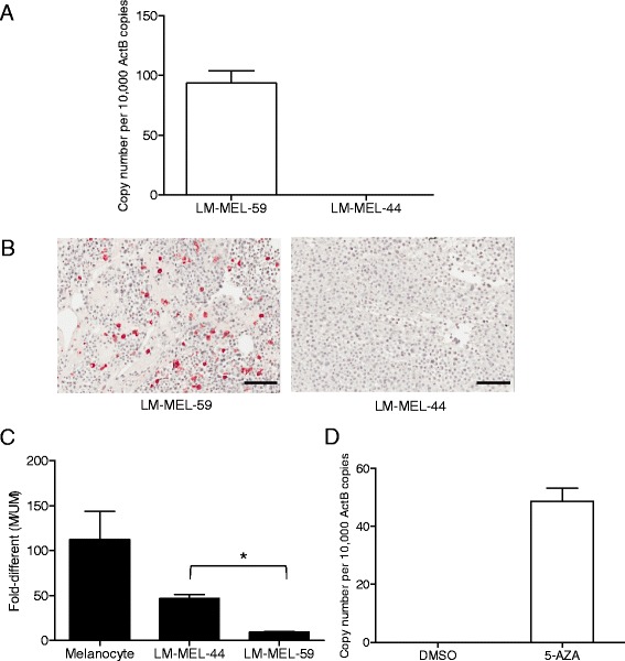
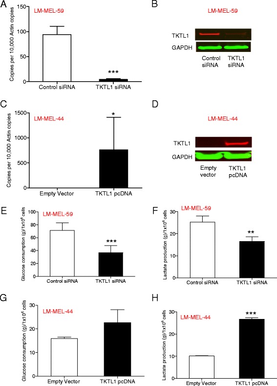
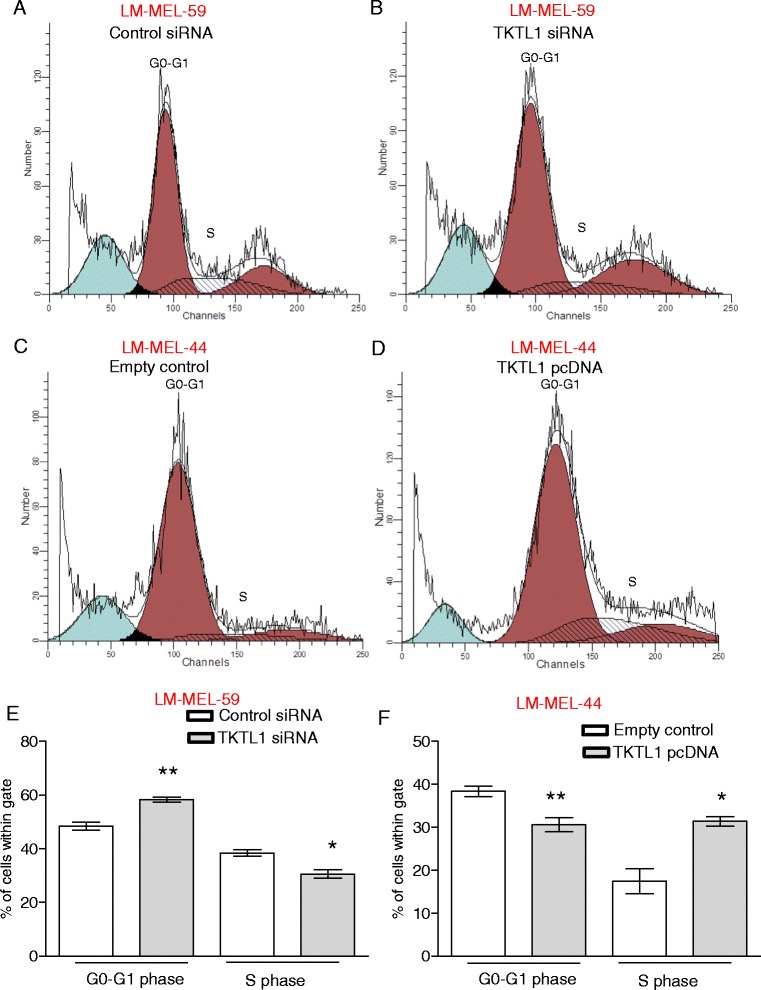
Results: Expression of TKTL1 was highly restricted in normal adult tissues and was overexpressed in a subset of metastatic melanoma tumors and derived cell lines. The TKTL1 promoter was activated by hypomethylation and treatment with 5aza induced TKTL1 expression in melanoma cells. Augmented expression of TKTL1 in melanoma cells was associated with a glycolytic phenotype. Loss and gain of function studies revealed that TKTL1 contributed to enhanced invasion of melanoma cells.

Conclusions: Our data provide evidence for an important role of TKTL1 in aerobic glycolysis and tumor promotion in melanoma that may result from defective promoter methylation. This epigenetic change may enable the natural selection of tumor cells with a metabolic phenotype and thereby provide a potential therapeutic target for a subset of melanoma tumors with elevated TKTL1 expression.


Figures

Fig. 1
Fig. 1
TKTL1 is highly expressed in human testis and melanoma tumors. a qRT-PCR was employed to measure the expression of TKTL1 in a panel of normal human tissues and in 38 metastatic melanoma tumor samples. b TKTL1 immunohistochemical staining in testis as positive control and control IgG staining in tumors as negative control are demonstrated. Representative staining patterns for TKTL1 in metastatic melanoma tumors are shown. Original magnification, 10 ×. c Graph shows number of TKTL1 positive and negative tumors
Fig. 2
Fig. 2
TKTL1 is expressed in metastatic melanoma cell lines and is regulated by promoter hypomethylation. a Using qRT-PCR the expression of TKTL1 in two melanoma cell lines, LM-MEL-59 and LM-MEL-44 was determined. b TKTL1 expression in LM-MEL-59 and LM-MEL-44 was determined by IHC. Original magnification, 20×. c Bisulfite conversion and MS-qPCR was performed to assay the promoter methylation status of TKTL1 in melanocytes and metastatic melanoma cells. Relative levels of methylated and unmethylated products (M/UM) was quantified. d QRT-PCR was employed to detect changes in expression of TKTL1 by 5-azacytidine treatment in LM-MEL-44 cell line. Values are ± SD of three independent experiments in triplicate (*, p < 0.05)
Fig. 3
Fig. 3
Ectopic overexpression and knockdown of TKTL1 influences the Warburg effect in melanoma cells. a QRT-PCR was employed to evaluate the expression of TKTL1 in LM-MEL-59 after treatment with either TKTL1 or control siRNA for 72 h. b Western Blotting with a mouse monoclonal anti-TKTL1 antibody showed reduction in TKTL1 levels after siRNA treatment in LM-MEL-59 after 72 h. GAPDH was used a loading control. c TKTL1 expression was assessed by qRT-PCR in LM-MEL-44 cells transfected with TKTL1 pcDNA or empty control for 72 h. d Immunoblot of TKTL1 confirmed expression of TKTL1 after transfection of a TKTL1 expression vector in LM-MEL-44. Blots were probed with GAPDH as a control for loading and transfer. Glucose consumption was measured in cell free supernatant of e LM-MEL-59 cell line following treatment with TKTL1 or control siRNA for 72 h and g LM-MEL-44 cell line following over-expression of TKTL1 or empty vector control for 72 h. The production of lactate in culture supernatants was measured in f LM-MEL-59 after knockdown of TKTL1 for 72 h and h LM-MEL-44 after treatment with TKTL1 pcDNA or empty vector control for 72 h. Values are ± SD of three experiments in triplicate (**, p < 0.005, ***, p < 0.0005)
Fig. 4
Fig. 4
Loss of TKTL1 expression changes cell cycle distribution of melanoma cells. Cell cycle phases were determined by propidium iodide staining of melanoma cells and subsequent flow cytometric analysis. A representative histogram of cell cycle analysis of LM-MEL-59 is shown after a control siRNA treatment and b TKTL1 siRNA treatment. Analysis of percentage of cells in e G0-G1 phase and S phase cell after treatment of LM-MEL-59 with TKTL1 or control siRNA. Values are ± SD of three experiments in triplicate (*, p < 0.05, **, p < 0.005). Histograms depicting distribution of cell cycle phase in LM-MEL-44 after treatment with c empty vector control and d TKTL1 pcDNA. Cell cycle distribution of f GO-G1 and S phase cell population after 48 h of ectopic expression of TKTL1 or empty vector in LM-MEL-44 was performed. Values are ± SD of three experiments in triplicate (*, p < 0.05, **, p < 0.005)
Fig. 5
Fig. 5
TKTL1 enhances invasive behaviour in melanoma. Melanoma cells were plated out and transfected with either 10nM control siRNA or TKTL1 specific siRNA and subjected to invasion assay. a Representative images of invasion of LM-MEL-59 is shown (scale bar = 100 μm). b The graph shows the total number of invasive cells counted. c Invasiveness of melanoma cells LM-MEL-44 after treatment with empty vector control or TKTL1 pcDNA was tested, images captured (scale bar = 100 μm) and d invasion was quantified as above. Values are mean ± SD of three independent experiments in triplicate (*p < 0.05, **p < 0.005)

References

    1. Little EG, Eide MJ. Update on the current state of melanoma incidence. Dermatol Clin. 2012;30(3):355–61. doi: 10.1016/j.det.2012.04.001. - DOI - PubMed
    1. Miller AJ, Mihm MC., Jr Melanoma. N Engl J Med. 2006;355(1):51–65. doi: 10.1056/NEJMra052166. - DOI - PubMed
    1. Finn L, Markovic SN, Joseph RW. Therapy for metastatic melanoma: the past, present, and future. BMC Med. 2012;10:23. doi: 10.1186/1741-7015-10-23. - DOI - PMC - PubMed
    1. Girotti MR, Saturno G, Lorigan P, Marais R. No longer an untreatable disease: How targeted and immunotherapies have changed the management of melanoma patients. Mol Oncol. 2014;8(6):1140-58. - PMC - PubMed
    1. Holderfield M, Deuker MM, McCormick F, McMahon M. Targeting RAF kinases for cancer therapy: BRAF-mutated melanoma and beyond. Nat Rev Cancer. 2014;14(7):455–67. doi: 10.1038/nrc3760. - DOI - PMC - PubMed

Publication types