. 2016 Feb 19;17(2):255.
doi: 10.3390/ijms17020255.

Prenylated Flavonoids from Cudrania tricuspidata Suppress Lipopolysaccharide-Induced Neuroinflammatory Activities in BV2 Microglial Cells

Affiliations

Prenylated Flavonoids from Cudrania tricuspidata Suppress Lipopolysaccharide-Induced Neuroinflammatory Activities in BV2 Microglial Cells

Dong-Cheol Kim et al. Int J Mol Sci. .

Abstract

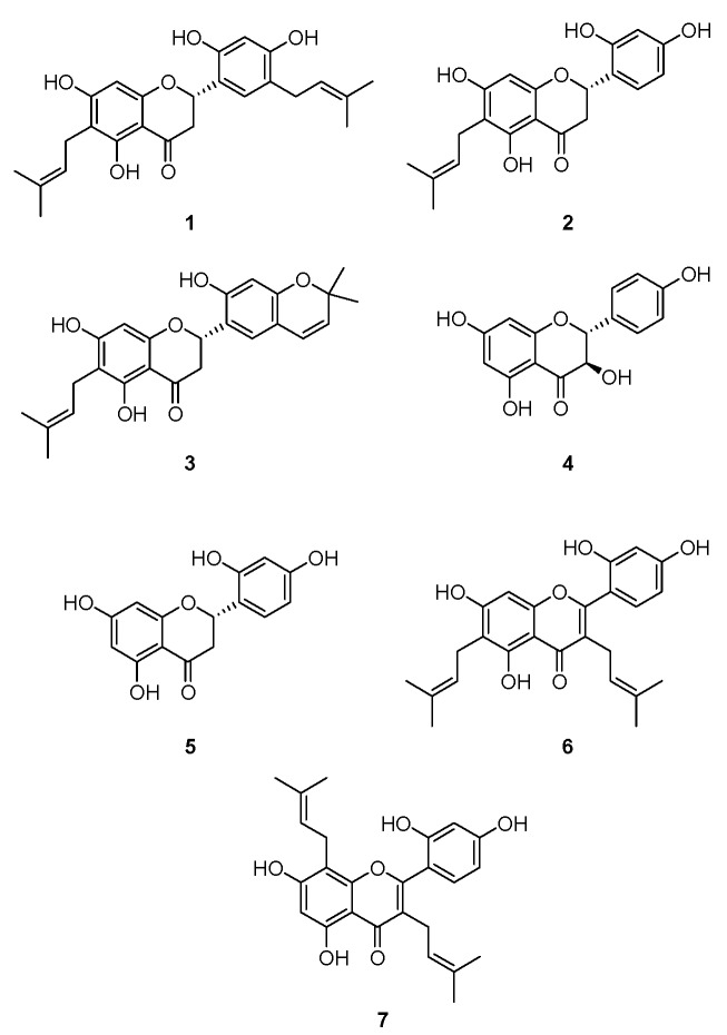
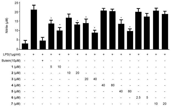
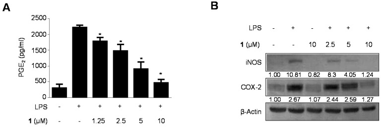
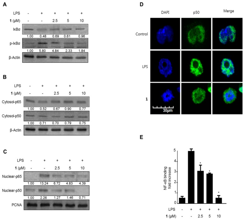
In Korea and China, Cudrania tricuspidata Bureau (Moraceae) is an important traditional medicinal plant used to treat lumbago, hemoptysis, and contusions. The C. tricuspidata methanol extract suppressed both production of NO and PGE₂ in BV2 microglial cells. Cudraflavanone D (1), isolated from this extract, remarkably suppressed the protein expression of inducible NO synthase and cyclooxygenase-2, and decreased the levels of NO and PGE₂ in BV2 microglial cells exposed to lipopolysaccharide. Cudraflavanone D (1) also decreased IL-6, TNF-α, IL-12, and IL-1β production, blocked nuclear translocation of NF-κB heterodimers (p50 and p65) by interrupting the degradation and phosphorylation of inhibitor of IκB-α, and inhibited NF-κB binding. In addition, cudraflavanone D (1) suppressed the phosphorylation of c-Jun N-terminal kinase (JNK) and p38 MAPK pathways. This study indicated that cudraflavanone D (1) can be a potential drug candidate for the cure of neuroinflammation.

Keywords: Cudrania tricuspidata; cudraflavanone D; microglia; mitogen-activated protein kinase; neuroinflammation; nuclear factor-κB.


Figures

Figure 1
Figure 1
The structures of compounds 17.
Figure 2
Figure 2
The effects of compounds 17 on nitrite production in BV2 microglial cells stimulated with LPS. The cells were pre-treated for 3 h with the indicated concentrations of compounds 17 and then stimulated for 24 h with LPS (1 μg/mL). The concentrations of nitrite were determined as described in the Materials and Methods section. The data represent the mean values ± SD of three experiments. * p < 0.05, as compared with cells treated with LPS only.
Figure 3
Figure 3
The effects of cudraflavanone D (1) on TNF-α (A), IL-1β (B), IL-12 (C), and IL-6 (D) mRNA expression in BV2 microglial cells stimulated with LPS. Cells were pre-treated for 3 h with the indicated concentrations of cudraflavanone D (1) and then stimulated for 12 h with LPS (1 μg/mL). The concentrations of TNF-α (A), IL-1β (B), IL-12 (C), and IL-6 (D) were determined as described in Materials and Methods. RNA quantification was performed as described in Materials and Methods and representative blots of three independent experiments are shown. The data represent the mean values of three experiments ± SD. * p < 0.05, as compared with the cells treated with LPS only.
Figure 3
Figure 3
The effects of cudraflavanone D (1) on TNF-α (A), IL-1β (B), IL-12 (C), and IL-6 (D) mRNA expression in BV2 microglial cells stimulated with LPS. Cells were pre-treated for 3 h with the indicated concentrations of cudraflavanone D (1) and then stimulated for 12 h with LPS (1 μg/mL). The concentrations of TNF-α (A), IL-1β (B), IL-12 (C), and IL-6 (D) were determined as described in Materials and Methods. RNA quantification was performed as described in Materials and Methods and representative blots of three independent experiments are shown. The data represent the mean values of three experiments ± SD. * p < 0.05, as compared with the cells treated with LPS only.
Figure 4
Figure 4
(A) The effects of cudraflavanone D (1) on protein expression of iNOS and COX-2 (B) in BV2 microglial cells stimulated with LPS. Cells were pre-treated for 3 h with the indicated concentrations of cudraflavanone D (1) and then stimulated for 24 h with LPS (1 μg/mL). The concentrations of iNOS and COX-2 (B) were determined as described in Materials and Methods. Western blot analyses were performed as described in Materials and Methods and representative blots of three independent experiments are shown. Band intensity was quantified by densitometry and normalized to β-actin; the values are presented below each band. Relative data represent the mean values of three experiments ± SD. * p < 0.05, as compared to the cells treated with LPS only.
Figure 5
Figure 5
The effects of cudraflavanone D (1) on IκB-α phosphorylation and degradation (A), NF-κB activation (B,C), NF-κB localization (D), and NF-κB DNA binding activity (E) in BV2 microglial cells. Cells were pre-treated for 3 h with the indicated concentrations of cudraflavanone D (1), and then stimulated for 1 h with LPS (1 μg/mL). Western blot analyses of IκB-α and phosphorylated (p)-IκB-α in the cytoplasm (A), and NF-κB in the cytoplasm (B) and nucleus (C), and immunofluorescent analysis (E), were performed as described in Materials and Methods. Band intensity was quantified by densitometry and normalized to β-actin and PCNA, and the values are presented below each band. Relative data represent the mean values of three experiments ± SD. * p < 0.05, as compared to the cells treated with LPS only.
Figure 6
Figure 6
The effects of cudraflavanone D (1) on ERK, JNK, and p38 MAPK protein expression and phosphorylation. Cells were pre-treated for 3 h with the indicated concentrations of cudraflavanone D (1) and stimulated for 1 h with LPS (1 μg/mL) (AC). The levels of (A) phosphorylated-ERK (p-ERK), (B) phosphorylated-JNK (p-JNK), and (C) phosphorylated-p38 MAPK (p-p38 MAPK) were determined by Western blotting. Representative blots from three independent experiments are shown. Band intensity was quantified by densitometry and normalized to β-actin; the values are presented below each band. Relative data represent the mean values of three experiments.

References

    1. Choi S.R., You D.H., Kim J.Y., Park C.B., Kim D.H., Ryu J. Antioxidant activity of methanol extracts from Cudrania tricuspidata Bureau according to harvesting parts and time. Korean J. Med. Crop. Sci. 2009;17:115–120.
    1. Shougakukan . The Dictionary of Chinese Drugs. Shanghai Science and Technological Publisher; Shanghai, China: 1985.
    1. Fujimoto T., Hano Y., Nomura T. Components of root bark of Cudrania tricuspidata l. 1,2 structures of four new isoprenylated xanthones, cudraxanthones A, B, C and D. Planta Med. 1984;50:218–221. doi: 10.1055/s-2007-969682. - DOI - PubMed
    1. Fujimoto T., Hano Y., Nomura T., Uzawa J. Components of root bark of Cudrania tricuspidata 2. Structures of two new isoprenylated flavones, cudraflavones A and B. Planta Med. 1984;50:161–163. doi: 10.1055/s-2007-969660. - DOI - PubMed
    1. Joo H.Y., Lim K.T. Glycoprotein isolated from Cudrania tricuspidata Bureau inhibits iNOS and COX-2 expression through modulation of NF-κB in LPS-stimulated RAW 264.7 cells. Environ. Toxicol. Pharmacol. 2009;27:247–252. doi: 10.1016/j.etap.2008.10.014. - DOI - PubMed

Publication types

MeSH terms

LinkOut - more resources