. 2016 Feb:38:16-23.
doi: 10.1016/j.placenta.2015.12.002. Epub 2015 Dec 13.

Abnormal labyrinthine zone in the Hectd1-null placenta

Affiliations

Abnormal labyrinthine zone in the Hectd1-null placenta

Anjali A Sarkar et al. Placenta. 2016 Feb.

Abstract

Introduction: The labyrinthine zone of the placenta is where exchange of nutrients and waste occurs between maternal and fetal circulations. Proper development of the placental labyrinth is essential for successful growth of the developing fetus and abnormalities in placental development are associated with intrauterine growth restriction (IUGR), preeclampsia and fetal demise. Our previous studies demonstrate that Hectd1 is essential for development of the junctional and labyrinthine zones of the placenta. Here we further characterize labyrinthine zone defects in the Hectd1 mutant placenta.

Methods: The structure of the mutant placenta was compared to wildtype littermates using histological methods. The expression of cell type specific markers was examined by immunohistochemistry and in situ hybridization.

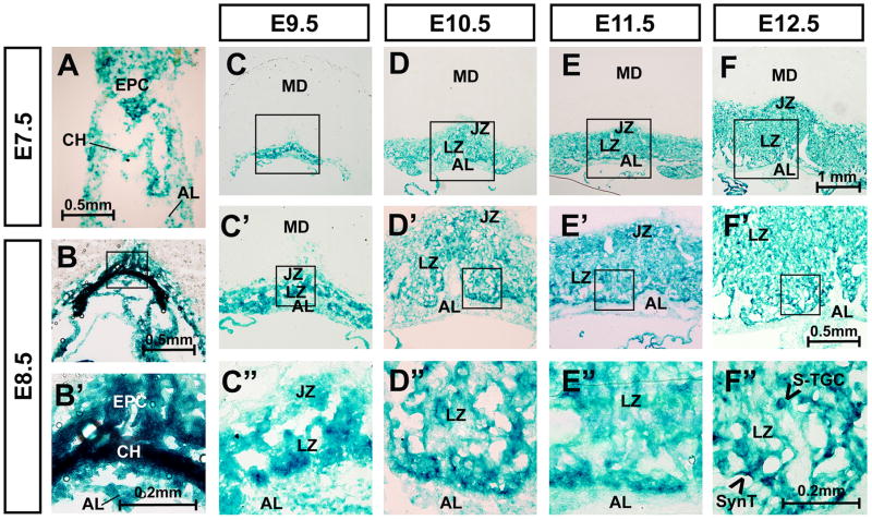
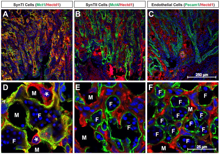
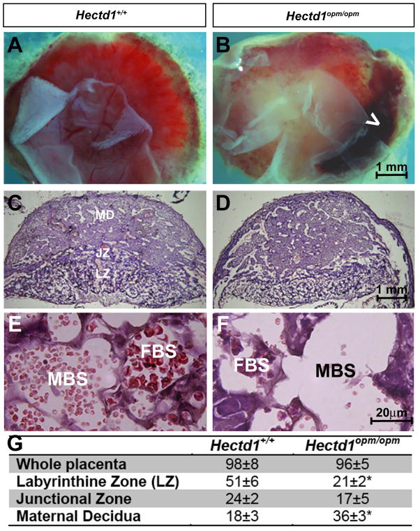
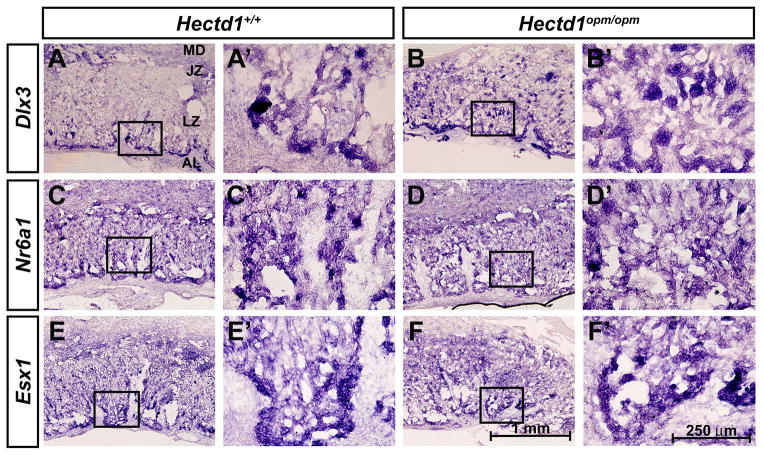
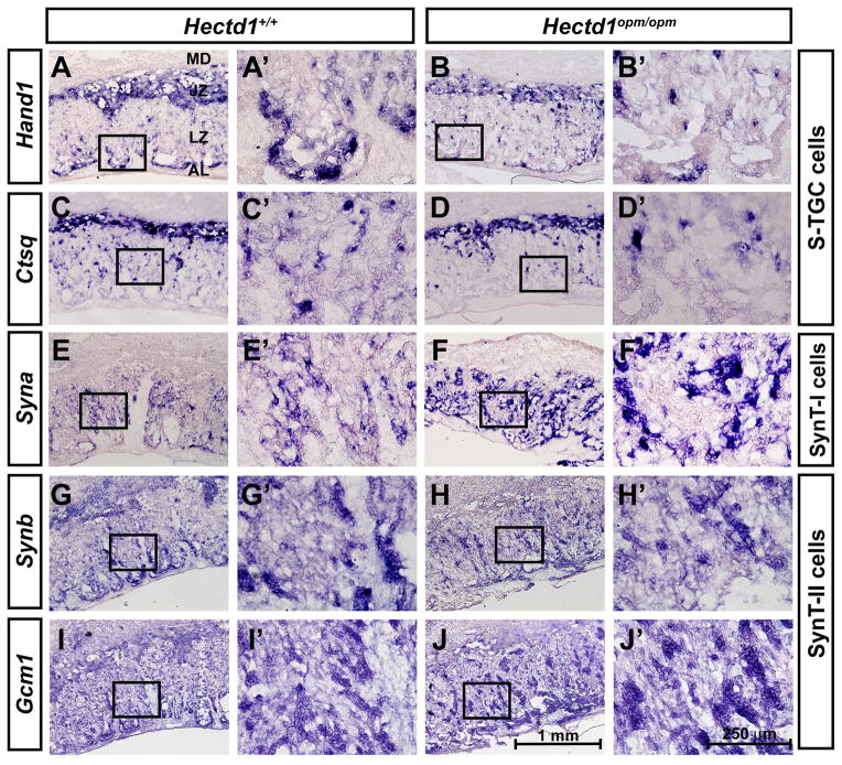
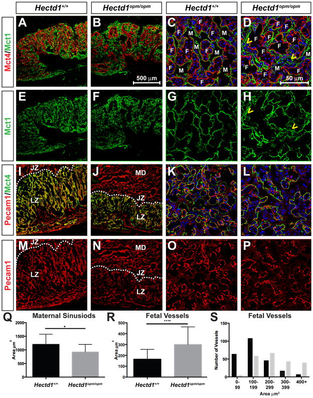
Results: Hectd1 is expressed in the labyrinthine zone throughout development and the protein is enriched in syncytiotrophoblast layer type I cells (SynT-I) and Sinusoidal Trophoblast Giant cells (S-TGCs) in the mature placenta. Mutation of Hectd1 results in pale placentas with frequent hemorrhages along with gross abnormalities in the structure of the labyrinthine zone including a smaller overall volume and a poorly elaborated fetal vasculature that contain fewer fetal blood cells. Examination of molecular markers of labyrinthine trophoblast cell types reveals increased Dlx3 positive cells and Syna positive SynT-I cells, along with decreased Hand1 and Ctsq positive sinusoidal trophoblast giant cells (S-TGCs).

Discussion: Together these defects indicate that Hectd1 is required for development of the labyrinthine zonethe mouse placenta.

Keywords: HECT E3 ligase; Labyrinthine layer; Placenta.


Figures

Figure 1
Figure 1. Hectd1 is expressed throughout development of the labyrinthine zone
A–F”) Hectd1 expression was monitored by LacZ staining in heterozygous Hectd1XC/+ placentas at E7.5, E8.5, 9.5, 10.5, 11.5 and E12.5 as indicated. Samples from crosses between wildtype females and Hectd1XC/+ males were used to label Hectd1 expressing cells derived from the conceptus and not the maternal decidua. Structures contributing to the placenta are labeled in E7.5 and 8.5 embryos including the ectoplacental cone (EPC), chorion (CH) and allantois (AL). The layers of the E9.5–12.5 placenta are labeled as maternal decidua (MD), junctional zone (JZ), labyrinthine zone (LZ) and allantoic mesoderm (AL). Magnification of boxed region is shown. LacZ staining is greater in the chorion and cells at the border of the chorionic-derived labyrinthine cells and allantoic mesoderm.
Figure 2
Figure 2. Hectd1 protein is localized to SynT-I and S-TGCs in the labyrinthine zone
Wildtype placentas were subjected to immunostaining with antibodies against Hectd1 along with Mct1 (A, D), Mct4 (B, E) or Pecam1 (C, F) to label SynT-I, SynT-II and endothelial cells respectively. Placentas in panels AC are transverse and D–F coronal sections. Hectd1 protein co-localizes with Mct1 but not Mct4 or Pecam1. Hectd1 protein is also found in more rounded Mct1-negative S-TGCs that line the maternal blood spaces (*). Examples of fetal vessels (F) and maternal blood spaces (M) are labeled.
Figure 3
Figure 3. Hemorrhages and altered histological appearance of Hectd1 mutant placentas
A, B) E12.5 (A) wildtype (Hectd1+/+) and (B) mutant (Hectd1opm/opm) placentas viewed from the fetal side demonstrating the overall pale appearance and large hemorrhages (white arrowhead) in the mutant. C, D) H&E staining of midsagittal section of E12.5 (C) wildtype and (D) mutant placentas. The layers of the placenta are labeled as maternal decidua (MD), junctional zone (JZ) and labyrinthine zone (LZ). E, F) Higher magnification images of the labyrinthine zone of H&E stained wildtype (E) and mutant (F) placentas. Maternal (MBS) and fetal blood spaces (FBS) are labeled that contain anucleated and nucleated red blood cells, respectively. G) Stereological analysis of Hectd1 wildtype and mutant placentas. Three placentas and wildtype siblings at E12.5 from three separate litters were utilized in all groups, mean values ± S.E.M, significant difference assessed by paired t-test between mutant and wildtype (* indicates p < 0.05).
Figure 4
Figure 4. Expression of labyrinthine trophoblast markers in the Hectd1 mutant placenta
In situ hybridization showing expression of Dlx3 (A, B), Nr6a1/Gcnf (C, D) and Esx1 (E, F) in E12.5 wildtype (A, C, E) and Hectd1opm/opm mutant (B, D, F) placentas. Images are shown at 10X and a 40X magnification of the boxed region.
Figure 5
Figure 5. Altered expression of cell-type specific labyrinthine trophoblast markers in the Hectd1 mutant placenta
Reduced Hand1 (A, B) and Ctsq (C, D) expressing S-TGCs and expanded expression of Syna (E, F) or Synb (G, H) and Gcm1 (I, J) (SynT-I or SynT-II cells, respectively) in E12.5 wildtype (A, C, E, G, I) and Hectd1opm/opm mutant (B, D, F, H, J) placentas. Images are shown at 10X and a 40X magnification of the boxed region.
Figure 6
Figure 6. Altered organization of SynT-I, SynT-II and fetal blood vessels in the Hectd1 mutant placenta
Expression of Mct1 (SynT-I cells, green) and Mct4 (SynT-II cells, red) were examined by immunofluorescence confocal microscopy in E12.5 wildtype (A, C, E, G) and mutant (B, F, D, H) placentas. Expression of Mct4 (SynT-II cells, green) and Pecam1 (endothelial cells, red) in E12.5 wildtype (I, K, M, O) and mutant (J, L, N, P) placentas. Nuclei are stained with Hoechst and shown in Blue in panels C, D, K and L. Placentas in panels A, B, E, F, I, J, M and N are sectioned in the transverse plane and shown at 10X and C, D, G, H, K, L, O and P in the coronal plane and shown at 60X. Yellow arrowheads in D and H highlight abnormal organization of SynT-I cells in the mutant. Fetal (F) and maternal (M) blood spaces are labeled. In panels I, J, M, N, the layers of the placenta are labeled as maternal decidua (MD), junctional zone (JZ) and labyrinthine zone. The dotted line denotes the boundary between junctional and labyrinthine zones. Q–S) The area of maternal (Q) and fetal (R, S) blood spaces were measured and the average area plotted (Q, R). 208 maternal blood spaces were measured in 13 wildtype and 155 blood spaces were measured in mutant sections from 4 wildtype and 2 mutant placentas. 237 fetal vessels in 12 wildtype and 210 fetal vessels in 18 mutant sections were measured from 4 wildtype and 2 mutant placentas. Significant differences were assessed by paired t-test between mutant and wildtype (* indicates p < 0.05 and *** p < 0.001). (S) The number of fetal vessels with the indicated area for wildtype (black bars) and Hectd1 mutant (grey bars).

Similar articles

Cited by

References

    1. Cross JC, Nakano H, Natale DR, Simmons DG, Watson ED. Branching morphogenesis during development of placental villi. Differentiation; research in biological diversity. 2006;74(7):393–401. - PubMed
    1. Sarkar AA, Nuwayhid SJ, Maynard T, Ghandchi F, Hill JT, Lamantia AS, Zohn IE. Hectd1 is required for development of the junctional zone of the placenta. Dev Biol. 2014;392(2):368–80. - PMC - PubMed
    1. Zohn IE, Anderson KV, Niswander L. The Hectd1 ubiquitin ligase is required for development of the head mesenchyme and neural tube closure. Developmental biology. 2007;306(1):208–21. - PMC - PubMed
    1. Sarkar AA, Zohn IE. Hectd1 regulates intracellular localization and secretion of Hsp90 to control cellular behavior of the cranial mesenchyme. The Journal of cell biology. 2012;196(6):789–800. - PMC - PubMed
    1. Nagy A. Manipulating the mouse embryo : a laboratory manual 2003. Cold Spring Harbor, N.Y: Cold Spring Harbor Laboratory Press; p. x.p. 764.

Publication types

Substances

LinkOut - more resources