. 2016 Apr 15;291(16):8414-27.
doi: 10.1074/jbc.M116.720201. Epub 2016 Feb 23.

Impaired Lysosomal Integral Membrane Protein 2-dependent Peroxiredoxin 6 Delivery to Lamellar Bodies Accounts for Altered Alveolar Phospholipid Content in Adaptor Protein-3-deficient pearl Mice

Affiliations

Impaired Lysosomal Integral Membrane Protein 2-dependent Peroxiredoxin 6 Delivery to Lamellar Bodies Accounts for Altered Alveolar Phospholipid Content in Adaptor Protein-3-deficient pearl Mice

Seunghyi Kook et al. J Biol Chem. .

Abstract

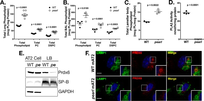
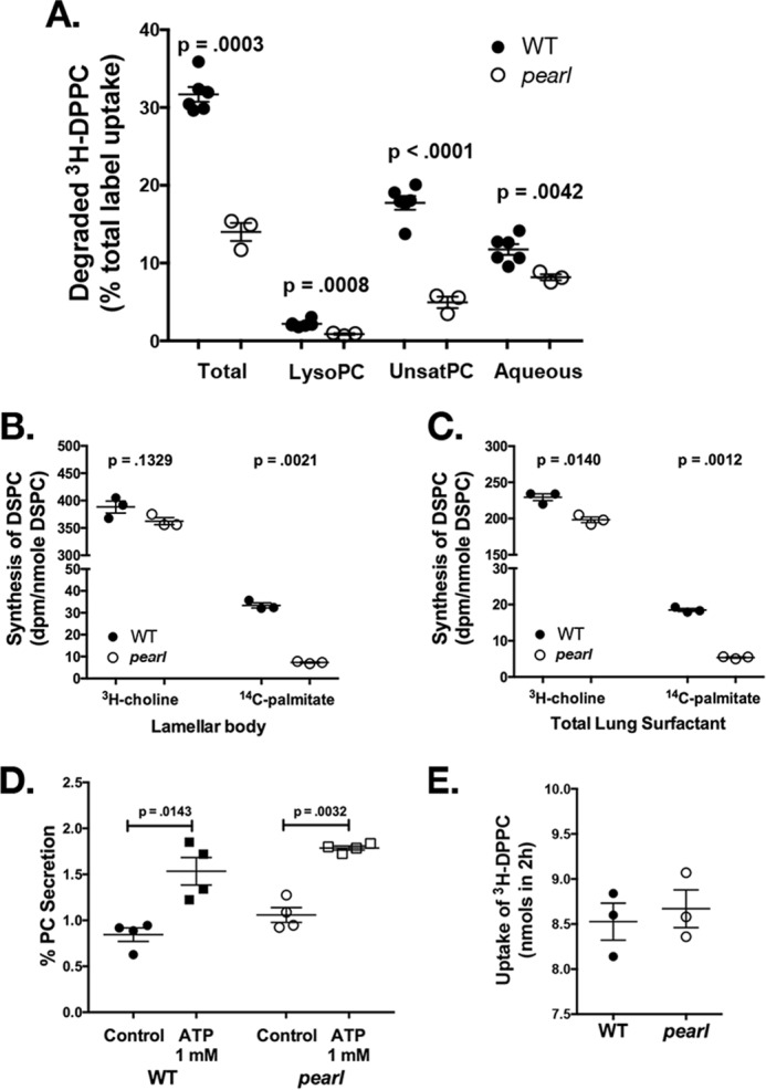
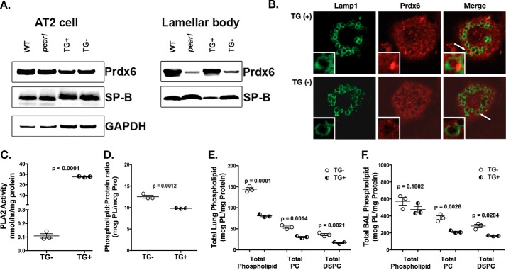
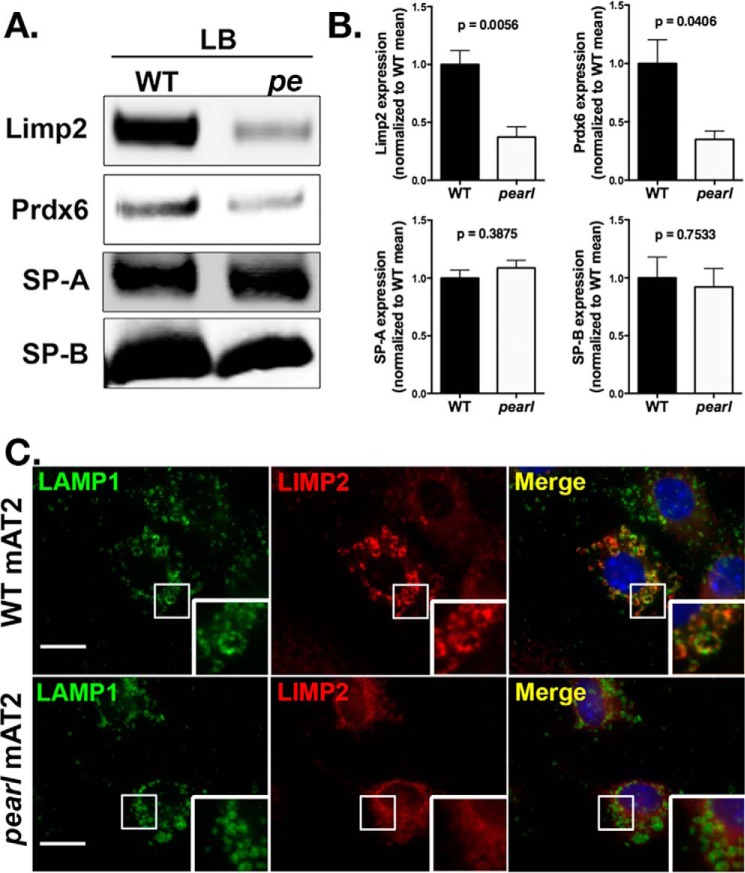
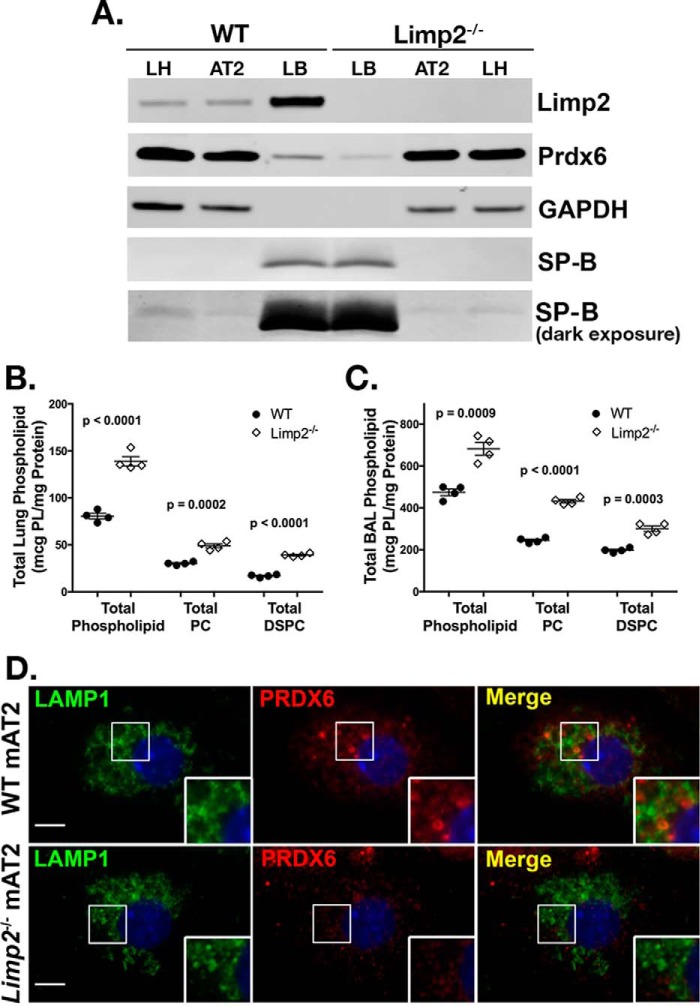
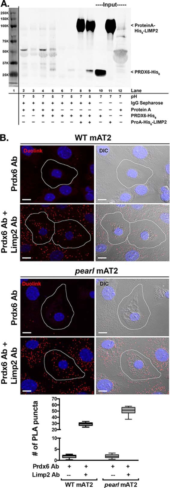
The Hermansky Pudlak syndromes (HPS) constitute a family of disorders characterized by oculocutaneous albinism and bleeding diathesis, often associated with lethal lung fibrosis. HPS results from mutations in genes of membrane trafficking complexes that facilitate delivery of cargo to lysosome-related organelles. Among the affected lysosome-related organelles are lamellar bodies (LB) within alveolar type 2 cells (AT2) in which surfactant components are assembled, modified, and stored. AT2 from HPS patients and mouse models of HPS exhibit enlarged LB with increased phospholipid content, but the mechanism underlying these defects is unknown. We now show that AT2 in the pearl mouse model of HPS type 2 lacking the adaptor protein 3 complex (AP-3) fails to accumulate the soluble enzyme peroxiredoxin 6 (PRDX6) in LB. This defect reflects impaired AP-3-dependent trafficking of PRDX6 to LB, because pearl mouse AT2 cells harbor a normal total PRDX6 content. AP-3-dependent targeting of PRDX6 to LB requires the transmembrane protein LIMP-2/SCARB2, a known AP-3-dependent cargo protein that functions as a carrier for lysosomal proteins in other cell types. Depletion of LB PRDX6 in AP-3- or LIMP-2/SCARB2-deficient mice correlates with phospholipid accumulation in lamellar bodies and with defective intraluminal degradation of LB disaturated phosphatidylcholine. Furthermore, AP-3-dependent LB targeting is facilitated by protein/protein interaction between LIMP-2/SCARB2 and PRDX6 in vitro and in vivo Our data provide the first evidence for an AP-3-dependent cargo protein required for the maturation of LB in AT2 and suggest that the loss of PRDX6 activity contributes to the pathogenic changes in LB phospholipid homeostasis found HPS2 patients.

Keywords: AP-3; Hermansky Pudlak syndrome; adaptor protein; alveolar epithelial type 2 cell; lamellar body; lung; peroxiredoxin; peroxiredoxin 6; phospholipid metabolism; trafficking.


Figures

FIGURE 1.
FIGURE 1.
Disrupted lamellar body phospholipid content in pearl mice is associated with loss of lamellar body PRDX6. A, total phospholipid, total PC, andtotal DSPC measured in lung tissue homogenate (n = 6 mice per genotype, mean ± S.E.). B, total phospholipid, total PC, and total DSPC measured in bronchoalveolar lavage fluid from wild type (WT) and pearl mice (n = 4 mice per genotype, mean ± S.E.). C, total phospholipid content measured in lamellar body fractions from WT and pearl mice (n = 3 samples per genotype, each prepared from three mice, mean ± S.E.). D, phospholipase A2 activity measured in lamellar body fractions isolated from WT and pearl mice (n = 4 samples from 2 to 3 mice of each strain, mean ± S.E.). E, representative immunoblots of PRDX6, SP-B, and GAPDH using lamellar body fractions (LB; 10 μg of protein) and alveolar type 2 cell lysate (AT2; 30 μg of protein) from wild type (WT) and pearl (pe) mice. F, immunolocalization of PRDX6 in WT and pearl isolated alveolar type 2 cells (mAT2) after depletion of cytosolic constituents, using LAMP1 immunostaining to identify lamellar bodies (representative of two experiments; bars, 5 μm).
FIGURE 2.
FIGURE 2.
Disrupted phospholipid homeostasis in pearl mice is associated with reduced DPPC degradation and DPPC synthesis. A, degradation of intratracheally instilled [3H]DPPC and recovery of metabolites at 2 h by isolated perfused lungs of WT and pearl mice. The result for each fraction (lyso-PC, unsaturated phosphatidylcholine, aqueous) is expressed as a percentage of total recovered radioactivity, which is the sum of the recoveries for these three fractions (n = 6 WT and 3 pearl mice; mean ± S.E.). B and C, incorporation of radiolabeled [3H]choline and [14C]palmitate into DSPC of lamellar bodies (B) and total lung surfactant (C) isolated from WT and pearl mouse lungs. Substrates were administered via tail vein injection 24 h prior to isolation of DSPC from lamellar body fractions and total lung homogenate. DSPC isolated from these samples was analyzed for radioactivity and total phosphorus. Synthesis was expressed as disintegrations/min per nmol of DSPC (n = 3; mean ± S.E.). D, secretion of (methyl-3H)-choline-labeled phospholipid from AT2 cells isolated from WT and pearl mice, calculated as a percentage of recovered disintegrations/min associated with phospholipid in the culture medium after 2 h of secretagogues divided by total disintegrations/min in the cells plus culture medium (n = 4 experiments, mean ± S.E.). E, uptake of intratracheally instilled [3H]DPPC by isolated perfused lungs of WT and pearl mice between 5 and 120 min of lung perfusion (n = 3; mean ± S.E.).
FIGURE 3.
FIGURE 3.
Reconstitution of AP-3 function in AT2 cells restores lamellar body PRDX6 and normalizes lung phospholipid homeostasis in pearl mice. pearl mice expressing the Ap3b1 transgene from an SP-C promoter (TG+) and transgene negative (TG−) littermates were compared with WT and genetically unmanipulated pearl mice (8–10 weeks old). A, representative immunoblots for PRDX6, SP-B, and GAPDH using AT2 lysates (20 μg each) and lamellar body fractions (25 μg each) from WT, pearl, TG+, and TG− mice. B, immunolocalization of PRDX6 in TG+ and TG− isolated alveolar type 2 cells after depletion of cytosolic constituents, using LAMP1 immunostaining to identify lamellar bodies (representative of two experiments; lamellar body noted by white arrow). C, phospholipase A2 activity measured in lamellar bodies from TG− and TG+ mice (n = 3, mean ± S.E.). D, total phospholipid measured in lamellar bodies from TG− and TG+ mice (n = 3; mean ± S.E.). E and F, total phospholipid (PL), total PC, and total DSPC were measured in total lung tissue (E) and bronchoalveolar lavage fluid (F) from TG− and TG+ mice (n = 3 mice of each genotype, mean ± S.E.).
FIGURE 4.
FIGURE 4.
Reduced targeting of LIMP-2/SCARB2 to lamellar bodies in pearl mice. Representative immunoblots (A) with densitometry (B) for LIMP-2/SCARB2, PRDX6, SP-A, and SP-B using lamellar body fractions (LB; 5 μg of protein) from wild type (WT) and pearl (pe) mice (n = 4 samples prepared from 2 to 4 mice each; mean ± S.D.). C, immunolocalization of LIMP-2/SCARB2 with LAMP1 identifying lamellar bodies in isolated alveolar type 2 cells from WT and pearl mice (representative of two experiments; bars represent 5 μm).
FIGURE 5.
FIGURE 5.
Limp-2−/− mice exhibit reduced lamellar body PRDX6 and increased surfactant phospholipid pool sizes. A, representative immunoblots of LIMP2/SCARB2, PRDX6, SP-B, and GAPDH in total lung (LH), alveolar type 2 (AT2) cells, and lamellar body fractions (LB) from wild type (WT) and Limp-2−/− mice (10 μg of total protein per lane). A light exposure of the SP-B immunoblot is presented as a loading control for lamellar body lanes; darker exposures demonstrate SP-B in lung homogenate and AT2 cell samples. B and C, total phospholipid, total phosphatidylcholine, and total DSPC were measured in total lung tissue (B) and bronchoalveolar lavage fluid from WT and Limp-2−/− mice (C) (n = 4 mice of each genotype, mean ± S.E.). D, immunolocalization of PRDX6 in isolated alveolar type 2 cells (mAT2) from Limp-2−/− mice and wild type littermates (WT) after depletion of cytosolic constituents, using LAMP1 immunostaining to identify lamellar bodies (representative of two experiments; bars represent 5 μm).
FIGURE 6.
FIGURE 6.
Protein/protein interaction between LIMP2/SCARB2 and PRDX6 in vitro and in vivo. A, co-immunoprecipitation of recombinant PRDX6-His6 by recombinant protein A-His6-LIMP2 is favored in buffer of pH 5 compared with buffer of pH 7 (lanes 8 and 9). Representative immunoblot from two separate experiments. Lane 1 demonstrates molecular weight markers visualized by scanning at 700 nm. Lanes 2–7 illustrate nonspecific interactions between IgG-Sepharose beads with protein A alone (lanes 2 and 3), protein A and PRDX6-His6 (lanes 4 and 5), or PRDX6-His6 alone (lanes 6 and 7), at pH 7 and 5. Lanes 10–12 contain input protein for PRDX6-His6 and protein A-His6-LIMP2. B, representative confocal images of the Duolink PLA interaction between mouse anti-PRDX6 and rabbit anti-LIMP2 (red puncta). For negative controls, mAT2 cells were incubated with mouse anti-PRDX6 alone. The nuclei are labeled with DAPI and differential interference contrast (DIC) images were captured to distinguish the lamellar bodies within mAT2 cells. The red puncta represent positive Duolink signals showing the interaction between PRDX6 and LIMP2; scale bars, 10 μm. Summary data are shown for average number of PLA products per AT2 cell and were derived from two separate experiments, each using lung epithelial cells isolated from four mice of each genotype with 20–25 single AT2 cell images analyzed per genotype per experiment for each of the conditions shown. The box and whiskers plot shows the median (center line), 75th and 25th percentiles (upper and lower limits of the box), and range of raw data (maximum to minimum as hinges). The data were analyzed by unpaired t test, which showed p < 0.0001 WT versus pearl using both antisera in the proximity ligation assay.

Similar articles

Cited by

References

    1. Whitsett J. A., Wert S. E., and Weaver T. E. (2015) Diseases of pulmonary surfactant homeostasis. Annu. Rev. Pathol. 10, 371–393 - PMC - PubMed
    1. Ridsdale R., Na C.-L., Xu Y., Greis K. D., and Weaver T. (2011) Comparative proteomic analysis of lung lamellar bodies and lysosome-related organelles. PLoS ONE 6, e16482. - PMC - PubMed
    1. Wei M. L. (2006) Hermansky-Pudlak syndrome: a disease of protein trafficking and organelle function. Pigment Cell Res. 19, 19–42 - PubMed
    1. Raposo G., and Marks M. S. (2007) Melanosomes–dark organelles enlighten endosomal membrane transport. Nat. Rev. Mol. Cell Biol. 8, 786–797 - PMC - PubMed
    1. Marks M. S., Heijnen H. F., and Raposo G. (2013) Lysosome-related organelles: unusual compartments become mainstream. Curr. Opin. Cell Biol. 25, 495–505 - PMC - PubMed

Publication types

MeSH terms

Supplementary concepts

LinkOut - more resources