

Unusual case of spinal epidermoid cyst and a concomitant spinal arachnoid cyst
- PMID: 26907820
- PMCID: PMC4769457
- DOI: 10.1136/bcr-2015-214002
Unusual case of spinal epidermoid cyst and a concomitant spinal arachnoid cyst
Abstract
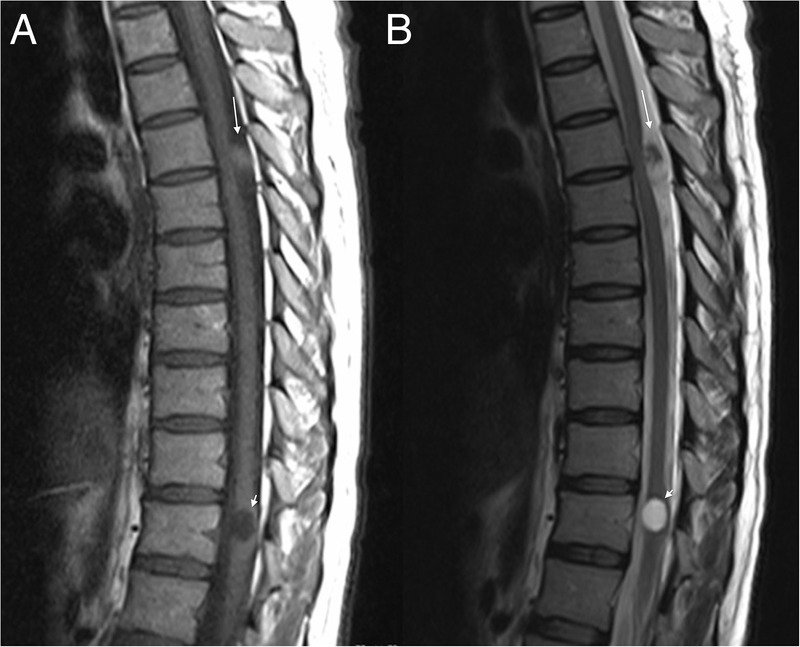
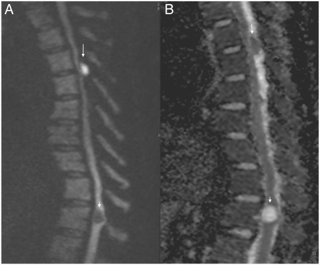
A 38-year-old woman presented with a 12-month history of subjective weakness and pain in her legs. Thoracolumbar MRI revealed two spinal intradural cystic lesions at T5-6 and T11 levels, respectively. The lesion located at the T5-6 level was heterogeneously hyperintense on T2-weighted images and heterogeneously hypointense on T1-weighted images. This lesion showed high signal intensity on diffusion weighted MRI (DWI) and low signal intensity on apparent diffusion coefficient images (ADC). According to the MRI findings, we reported this tumour as a spinal epidermoid cyst. The pathology result suggested that the lesion was an epidermoid cyst. The second intradural lesion, at the T11 level, showed a hypointense signal on T1 and hyperintense signal on T2 images. However, in contrast to the superior lesion, this lesion was hypointense on DWI and hyperintense on ADC. We evaluated the second lesion as an arachnoid cyst according to the MRI findings.
2016 BMJ Publishing Group Ltd.
Figures
References
Publication types
MeSH terms
Supplementary concepts
LinkOut - more resources
Full Text Sources
Other Literature Sources
Medical