The Value of Diffusion-Weighted Imaging in the Differential Diagnosis of Ovarian Lesions: A Meta-Analysis
- PMID: 26907919
- PMCID: PMC4764370
- DOI: 10.1371/journal.pone.0149465
The Value of Diffusion-Weighted Imaging in the Differential Diagnosis of Ovarian Lesions: A Meta-Analysis
Abstract
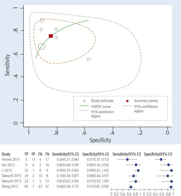
Objectives: The ability of contrast-enhanced MRI to distinguish between malignant and benign ovarian masses is limited. The aim of this meta-analysis is to evaluate the diagnostic performance of diffusion-weighted imaging (DWI) in differentiating malignant from benign ovarian masses.
Methods: A comprehensive literature search was performed in several authoritative databases to identify relevant articles. The weighted mean difference (WMD) and corresponding 95% confidence interval (95% CI) were calculated. We also used subgroup analysis to analyze study heterogeneity, and evaluated publication bias.
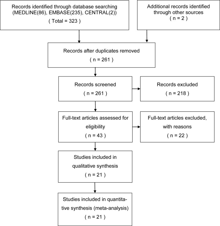
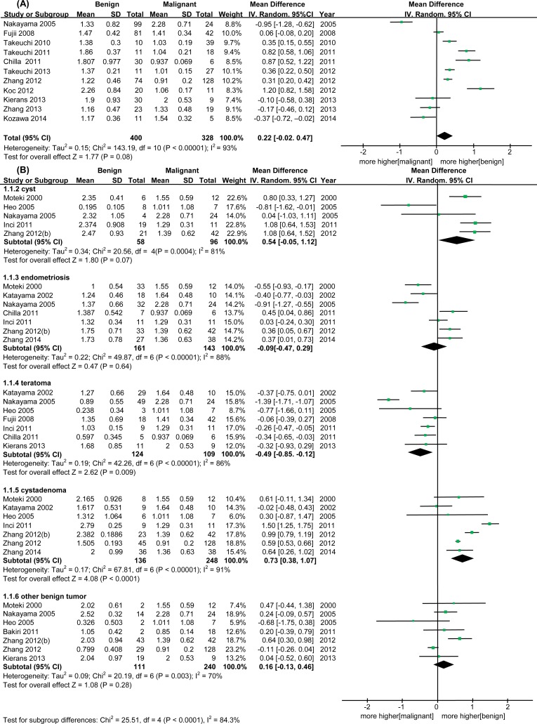
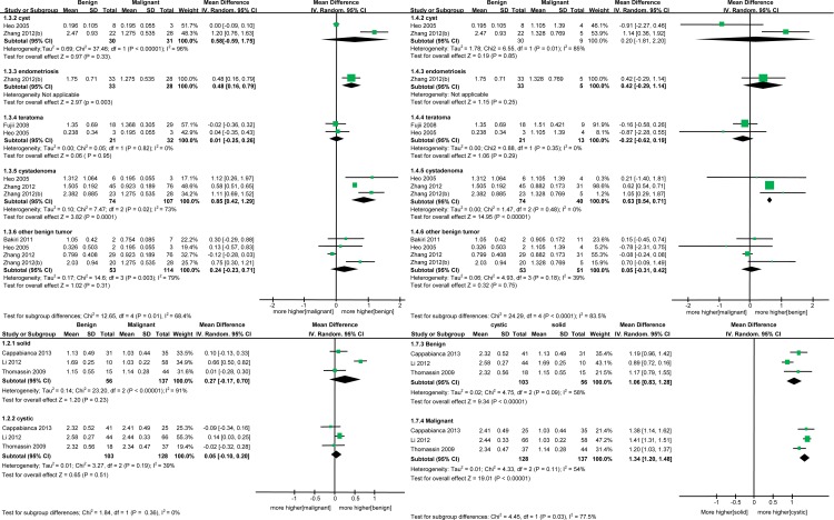
Results: The meta-analysis is based on 21 studies, which reported the findings for 731 malignant and 918 benign ovarian masses. There was no significant difference in apparent diffusion coefficient (ADC) values for DWI between benign and malignant lesions (WMD = 0.22, 95% CI = -0.02-0.47, p = 0.08). Subgroup analysis by benign tumor type revealed higher ADC values (or a trend toward higher values) for cysts, cystadenomas and other benign tumors compared to malignant masses (cyst: WMD = 0.54, 95% CI = -0.05-1.12, p = 0.07; cystadenoma: WMD = 0.73, 95% CI = 0.38-1.07, p < 0.0001; other benign tumor: WMD = 0.16, 95% CI = -0.13-0.46, p = 0.28). On the other hand, lower ADC values (or a trend toward lower values) were observed for endometrioma and teratoma compared to malignant masses (endometrioma: WMD = -0.09, 95% CI = -0.47-0.29, p = 0.64; teratoma: WMD = -0.49, 95% CI = -0.85-0.12, p = 0.009). Subgroup analysis by mass property revealed higher ADC values in cystic tumor types than in solid types for both benign and malignant tumors. Significant study heterogeneity was observed. There was no notable publication bias.
Conclusions: Quantitative DWI is not a reliable diagnostic method for differentiation between benign and malignant ovarian masses. This knowledge is essential in avoiding misdiagnosis of ovarian masses.
Conflict of interest statement
Figures


Similar articles
-
Diffusion-weighted MRI: a useful technique to discriminate benign versus malignant ovarian surface epithelial tumors with solid and cystic components.Abdom Imaging. 2012 Oct;37(5):897-903. doi: 10.1007/s00261-011-9814-x. Abdom Imaging. 2012. PMID: 22038329
-
Diffusion-weighted echo-planar MR imaging and ADC mapping in the differential diagnosis of ovarian cystic masses: usefulness of detecting keratinoid substances in mature cystic teratomas.J Magn Reson Imaging. 2005 Aug;22(2):271-8. doi: 10.1002/jmri.20369. J Magn Reson Imaging. 2005. PMID: 16028258
-
Endometrioid adenocarcinoma of the ovary: MRI findings with emphasis on diffusion-weighted imaging for the differentiation of ovarian tumors.Acta Radiol. 2016 Jun;57(6):758-66. doi: 10.1177/0284185115599805. Epub 2015 Aug 24. Acta Radiol. 2016. PMID: 26307063
-
MR imaging in discriminating between benign and malignant paediatric ovarian masses: a systematic review.Eur Radiol. 2020 Feb;30(2):1166-1181. doi: 10.1007/s00330-019-06420-4. Epub 2019 Sep 16. Eur Radiol. 2020. PMID: 31529256 Free PMC article.
-
Diagnostic accuracy of apparent diffusion coefficient (ADC) value in differentiating malignant from benign solid liver lesions: a systematic review and meta-analysis.Br J Radiol. 2021 Jul 1;94(1123):20210059. doi: 10.1259/bjr.20210059. Epub 2021 Jun 16. Br J Radiol. 2021. PMID: 34111960 Free PMC article.
Cited by
-
Differentiation of Endometriomas from Ovarian Hemorrhagic Cysts at Magnetic Resonance: The Role of Texture Analysis.Medicina (Kaunas). 2020 Sep 23;56(10):487. doi: 10.3390/medicina56100487. Medicina (Kaunas). 2020. PMID: 32977428 Free PMC article.
-
Diffusion-Weighted Magnetic Resonance Imaging as a Noninvasive Parameter for Differentiating Benign and Malignant Intraperitoneal Collections.Medicina (Kaunas). 2020 May 1;56(5):217. doi: 10.3390/medicina56050217. Medicina (Kaunas). 2020. PMID: 32369983 Free PMC article.
-
Ovarian Sertoli-Leydig Tumour in a Postmenopausal Female With Virilisation: Magnetic Resonance Imaging Findings.Cureus. 2025 Jun 13;17(6):e85965. doi: 10.7759/cureus.85965. eCollection 2025 Jun. Cureus. 2025. PMID: 40662027 Free PMC article.
-
Radiomic Analysis of MRI Images is Instrumental to the Stratification of Ovarian Cysts.J Pers Med. 2020 Sep 14;10(3):127. doi: 10.3390/jpm10030127. J Pers Med. 2020. PMID: 32937851 Free PMC article.
-
Apparent diffusion coefficient histogram analysis for differentiating solid ovarian tumors.Front Oncol. 2022 Aug 1;12:904323. doi: 10.3389/fonc.2022.904323. eCollection 2022. Front Oncol. 2022. PMID: 35978817 Free PMC article.
References
Publication types
MeSH terms
LinkOut - more resources
Full Text Sources
Other Literature Sources
Medical