Review
. 2016 May;33(5):1055-73.
doi: 10.1007/s11095-016-1885-5. Epub 2016 Feb 23.

Microneedles: A New Frontier in Nanomedicine Delivery

Affiliations
Review

Microneedles: A New Frontier in Nanomedicine Delivery

Eneko Larrañeta et al. Pharm Res. 2016 May.

Abstract

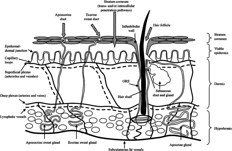
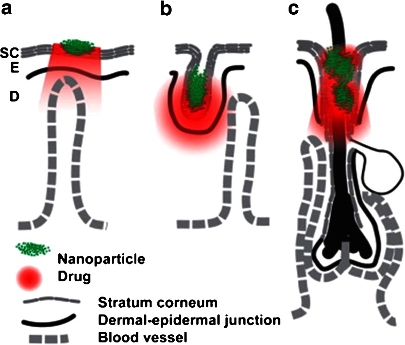
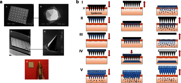
This review aims to concisely chart the development of two individual research fields, namely nanomedicines, with specific emphasis on nanoparticles (NP) and microparticles (MP), and microneedle (MN) technologies, which have, in the recent past, been exploited in combinatorial approaches for the efficient delivery of a variety of medicinal agents across the skin. This is an emerging and exciting area of pharmaceutical sciences research within the remit of transdermal drug delivery and as such will undoubtedly continue to grow with the emergence of new formulation and fabrication methodologies for particles and MN. Firstly, the fundamental aspects of skin architecture and structure are outlined, with particular reference to their influence on NP and MP penetration. Following on from this, a variety of different particles are described, as are the diverse range of MN modalities currently under development. The review concludes by highlighting some of the novel delivery systems which have been described in the literature exploiting these two approaches and directs the reader towards emerging uses for nanomedicines in combination with MN.

Keywords: drug delivery; microneedles; microparticles; nanomedicine; nanoparticles; vaccines.


Figures

Fig. 1
Fig. 1
Structures of different nanomedicines and their approximate sizes. For comparative purposes, the sizes of biological nanostructures are shown at the top of the figure. Reproduced with permission from the British Society For Nanomedicine.
Fig. 2
Fig. 2
Diagrammatic representation of skin structure. Reproduced with permission from (27).
Fig. 3
Fig. 3
Sites in skin for NP delivery. Topical NP drug delivery takes place in three major sites: SC surface (a), furrows (b), and openings of hair follicles (c). The NPs are depicted in green and the drug in red. Other sites for delivery are the viable epidermis (e) and dermis (d). Reproduced with permission from (4).
Fig. 4
Fig. 4
Images of a typical silicon MN arrays (a). Scanning electron microscopy (SEM) images taken of a typical silicon MN array (ad) and a digital photograph of a typical silicon MN array (e). Schematic representation of the main MN modalities (b). Solid MNs (I); coated MNs (II); dissolving MNs (III); hollow MNs (IV) and hydrogel-forming MNs (V). Reproduced with permission from (69).
Fig. 5
Fig. 5
Skin permeability to molecules and particles of different sizes after treatment with MN arrays (I). The permeability of human cadaver epidermis was increased by orders of magnitude with a 400-needle array inserted (□) and after the array was removed (•) for calcein, insulin, BSA, and latex nanospheres of 25 nm and 50 nm radius. Predictions are shown for needles inserted (dashed line) and needles removed (solid line) by using a mathematical model. Scanning electron micrographs of human epidermal membranes treated with a silicon MN device and a subsequent topical application of a fluorescent nanosphere formulation (II). (a and d), scale bar = 50 μm; (b and c), scale bar = 10 μm; (e and f), scale bar = 2 μm. Percentage of the applied dose of NPs deposited within the epidermis and dermis within a 48-h period (III). The skin without MN treatment was used in the control groups. Reproduced with permission from (75,106,107).
Fig. 6
Fig. 6
Fluorescence micrographs of histological sections after microinjection of 2.5 μm fluorescent microspheres into hairless rat skin in vivo under pressures ranging from 2.5 to 20 psi via the same needle and loading for the same time periods (a). Confocal microscopic images of 0.7 μm large fluorescent polystyrene microspheres at different depths in chicken tissue after injection with a MEMS syringe (b). Multi-layered MN inserted into pig cadaver skin, cryo-sectioned vertically and viewed by brightfield microscopy (c). Reproduced with permission from (96,111,112).
Fig. 7
Fig. 7
Schematic representation of the formation and triggered insulin delivery of the glucose responsive vesicles (a). Schematic of the release from integrated MN/glucose responsive vesicles patch mechanism of action (b). Reproduced with permission from (120).
Fig. 8
Fig. 8
Delivery of doxorubicin in porcine buccal tissue after insertion of MN arrays coated with PLGA NPs and loaded with doxorobucin. Confocal fluorescent microscopy images showing the distribution of the NPs after insertion of the arrays in the porcine buccal tissue. The bar graph shows variation in the mean fluorescence intensity as a function of depth from the insertion site. Scale bar represents: 100 μm. Reproduced with permission from (121).
Fig. 9
Fig. 9
Schematic representation of microneedle arrays penetrating the skin layers releasing nanoparticles, containing vaccine antigens. Antigens are processed by dendritic cells and antigen fragments are presented to T lymphocytes. T lymphocytes become active to CD4+ & CD8+ which then help to destroy tumour cells and viral pathogens.
Fig. 10
Fig. 10
Schematic representation of the mechanism of responsive failure of MN arrays containing hydrogel MPs (a). Schematic diagram of triggered transdermal drug delivery using near-infrared light-responsive MN (b). Reproduced with permission from (151,152).

References

    1. Wagner V, Dullaart A, Bock AK, Zweck A. The emerging nanomedicine landscape. Nat Biotechnol. 2006;24(10):1211–7. doi: 10.1038/nbt1006-1211. - DOI - PubMed
    1. Moghimi SM, Hunter AC, Murray JC. Nanomedicine: current status and future prospects. FASEB J. 2005;19(3):311–30. doi: 10.1096/fj.04-2747rev. - DOI - PubMed
    1. Parveen S, Misra R, Sahoo SK. Nanoparticles: a boon to drug delivery, therapeutics, diagnostics and imaging. Nanomedicine. 2012;8(2):147–66. - PubMed
    1. Prow TW, Grice JE, Lin LL, Faye R, Butler M, Becker W, et al. Nanoparticles and microparticles for skin drug delivery. Adv Drug Deliv Rev. 2011;63(6):470–91. doi: 10.1016/j.addr.2011.01.012. - DOI - PubMed
    1. Patravale V, Dandekar P, Jain R. Nanoparticulate drug delivery: perspectives on the transition from laboratory to market: Elsevier Science; 2012.

MeSH terms