. 2016 Feb 24:6:21522.
doi: 10.1038/srep21522.

Pulmonary Tuberculosis in Humanized Mice Infected with HIV-1

Affiliations

Pulmonary Tuberculosis in Humanized Mice Infected with HIV-1

Rebecca J Nusbaum et al. Sci Rep. .

Abstract

Co-infection with HIV increases the morbidity and mortality associated with tuberculosis due to multiple factors including a poorly understood microbial synergy. We developed a novel small animal model of co-infection in the humanized mouse to investigate how HIV infection disrupts pulmonary containment of Mtb. Following dual infection, HIV-infected cells were localized to sites of Mtb-driven inflammation and mycobacterial replication in the lung. Consistent with disease in human subjects, we observed increased mycobacterial burden, loss of granuloma structure, and increased progression of TB disease, due to HIV co-infection. Importantly, we observed an HIV-dependent pro-inflammatory cytokine signature (IL-1β, IL-6, TNFα, and IL-8), neutrophil accumulation, and greater lung pathology in the Mtb-co-infected lung. These results suggest that in the early stages of acute co-infection in the humanized mouse, infection with HIV exacerbates the pro-inflammatory response to pulmonary Mtb, leading to poorly formed granulomas, more severe lung pathology, and increased mycobacterial burden and dissemination.


Figures

Figure 1
Figure 1. HIV infection in HuMice co-infected with Mtb.
BLT HuMice were infected i.v. with 2,500 TCID50 of HIV-1 (JR-CSF) or mock infected (PBS). Subsets of HuMice from HIV or non-infected mice were infected i.n. with 250 CFU Mtb (H37Rv), 3 wk post-HIV infection. Data are means ± SEM from samples of non-infected control (n = 4) and three infection groups, HIV (n = 3), Mtb (n = 6) and Mtb/HIV (n = 8), of HuMice. (A) HIV p24 capsid protein was detected in HuMouse spleen and plasma by ELISA at 8 and 5 wk p.i. with HIV, and Mtb, respectively. Viral load did not differ among HIV and Mtb/HIV infection groups and results are thus pooled. (B) Flow cytometric detection of human CD45 + CD3 + CD4 + T cells in peripheral blood, shown as a percent of baseline (pre-HIV infection) value at 5 wk p.i. with Mtb.
Figure 2
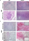
Figure 2. HIV-infected cells localize to Mtb lesions in the lung.
Animals were infected i.v. with 2500 TCID50 HIV-1 (JR-CSF) or mock infected and 3 wk later infected i.n. with 250 CFU of Mtb H37Rv. (A) Tissue pathology visualized by H&E staining shows normal lung architecture in a non-infected and HIV-infected animals and foci of inflammation in Mtb- and Mtb/HIV-infected lung. (B) Detection of HIV p24 by IHC (fast red) in lung of co-infected human reference samples at early and later stages of lesion progression, and in HIV- and HIV/Mtb-infected HuMice 8 and 5 wk p.i. with HIV and Mtb, respectively.
Figure 3
Figure 3. Pulmonary growth of Mtb is increased by HIV co-infection prior to peripheral CD4+ T cell depletion.
(A) CFU enumeration of Mtb in lung and liver tissue of HuMice at 5 wk p.i. with Mtb from two independent studies. Statistically significant differences compared to non-infected are designated by *p < 0.05. (B) Increased bacterial growth confirmed by Ziehl-Neelson detection of AFB by light microscopy in representative lung of Mtb-infected (left) and Mtb/HIV co-infected mice (right). (C) Correlation between HIV p24 and mycobacterial proliferation or pathology in the lung of co-infected HuMice (n = 8) was determined using a Spearman’s rank correlation co-efficient (Rs).
Figure 4
Figure 4. HIV co-infection exacerbates Mtb pathology in HuMouse lung.
Determination of pulmonary pathology using H&E visualization and light microscopy. (A) Representative lung from HuMouse 5 wk p.i. with Mtb or Mtb/HIV demonstrating increased number and/or size of TB lesions due to HIV. (B) Area of the lung, shown as a %, occupied by the average individual granuloma, or total granulomatous tissue, in tissue of HuMice infected with Mtb (n = 6) or Mtb/HIV (n = 8) as determined using Image J software. (C) Diffuse lesion development and poor containment of Mtb-infected macrophages due to HIV co-infection. (D) Increased pulmonary pathology and bacterial burden in necrotic tissue in co-infected animals. Insets show detection of AFB using Ziehl-Neelson staining in matched tissue sections. Statistically significant differences among treatment groups are designated as follows: *p < 0.05.
Figure 5
Figure 5. Co-infection with HIV promotes neutrophil accumulation in pulmonary TB granulomas.
(A) Visualization of lung pathology due to Mtb infection and Mtb/HIV co-infection in reference human (top), and representative humanized mouse (bottom) lung, demonstrating diffuse lesions and tissue damage. (B), Determination of neutrophil accumulation due to Mtb or Mtb/HIV infection through detection of myeloperoxidase (MPO) in infected human (top) and HuMouse lung (bottom) tissue. Top inset shows non-specific control staining for fast red in the necrotic lesion. Bottom inset shows representative tissue from HuMouse with HIV mono-infection, demonstrating lack a paucity of neutrophils and non-remarkable pathology in the absence of Mtb co-infection.
Figure 6
Figure 6. HIV co-infection activates excessive pro-inflammatory cytokine and chemokine responses to Mtb in the lung.
Lung supernatants from HuMice harvested 5 wk p.i. with Mtb or Mtb/HIV were used to determine activation of human cytokines and chemokines by multiplex ELISA. (A) Expression of human cytokines, and (B) chemokines, in non-, HIV-, Mtb-, and Mtb/HIV-infected HuMice. Data are means ± SEM from samples of non-infected control (n = 4) and three infection groups of Humice including HIV (n = 3), Mtb (n = 6) and Mtb/HIV (n = 8). Statistically significant differences compared to non-infected controls are designated as follows: *p < 0.05; **p < 0.01; ***p < 0.001. Significant differences among infected groups are shown as formula imagep < 0.05; formula imageformula imagep < 0.01;formula image formula image formula imagep < 0.001.
Figure 7
Figure 7. Pulmonary pneumonia and vascular occlusions develop as complications of HIV/Mtb co-infection.
Visualization of lung pathology due to Mtb infection and Mtb/HIV co-infection in representative HuMice (top) and reference human (bottom) lung. (A) Movat staining reveals vascular occlusion as indicated by blockage interior to elastin bands of vessel (black). Visualization of H&E staining demonstrates (B) TB pneumonia and (C) bronchial occlusions in co-infected HuMouse (top) and human (bottom) lung.

References

    1. Organization, W. H. (World Health Organization, Geneva, 2014).
    1. Harries A. D., Lawn S. D., Getahun H., Zachariah R. & Havlir D. V. HIV and tuberculosis–science and implementation to turn the tide and reduce deaths. J Int AIDS Soc 15, 17396 (2012). - PMC - PubMed
    1. Khan F. A. et al.. Treatment of active tuberculosis in HIV-coinfected patients: a systematic review and meta-analysis. Clin Infect Dis 50, 1288–99 (2010). - PubMed
    1. Law K. F., Jagirdar J., Weiden M. D., Bodkin M. & Rom W. N. Tuberculosis in HIV-positive patients: cellular response and immune activation in the lung. Am J Respir Crit Care Med 153, 1377–84 (1996). - PubMed
    1. Markowitz N. et al.. Incidence of tuberculosis in the United States among HIV-infected persons. The Pulmonary Complications of HIV Infection Study Group. Ann Intern Med 126, 123–32 (1997). - PubMed

Publication types