. 2016 May 15:132:425-438.
doi: 10.1016/j.neuroimage.2016.02.045. Epub 2016 Feb 22.

A multi-layer network approach to MEG connectivity analysis

Affiliations

A multi-layer network approach to MEG connectivity analysis

Matthew J Brookes et al. Neuroimage. .

Abstract

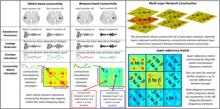
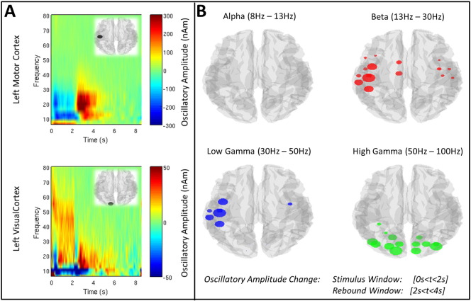
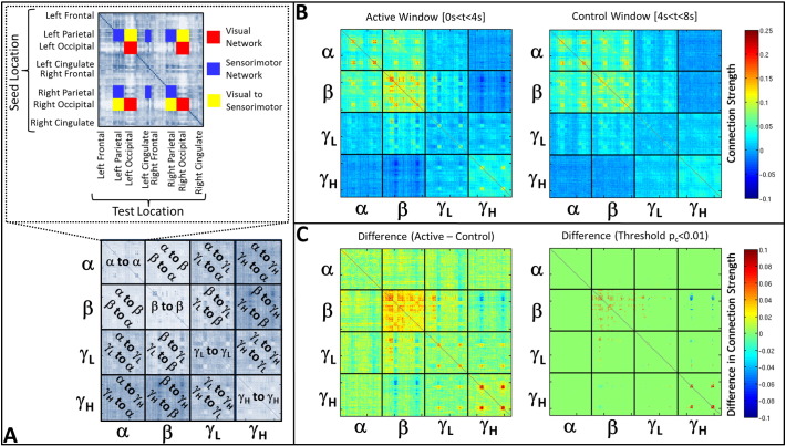
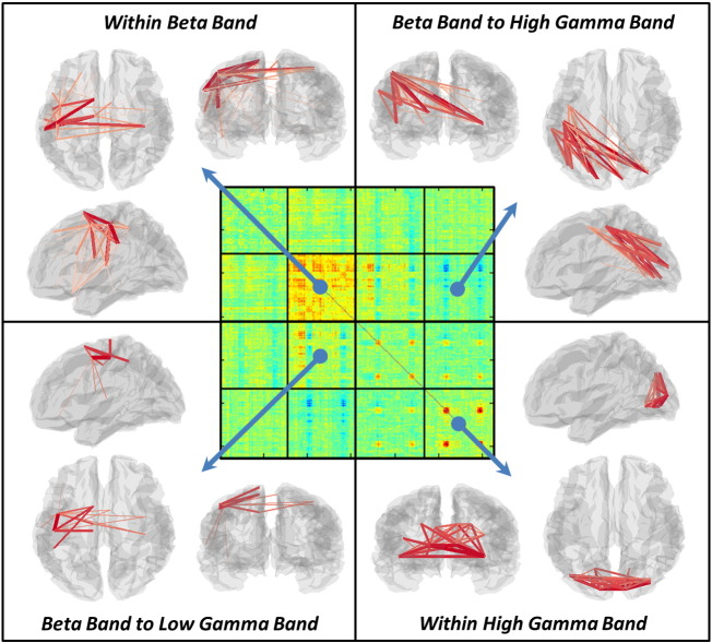
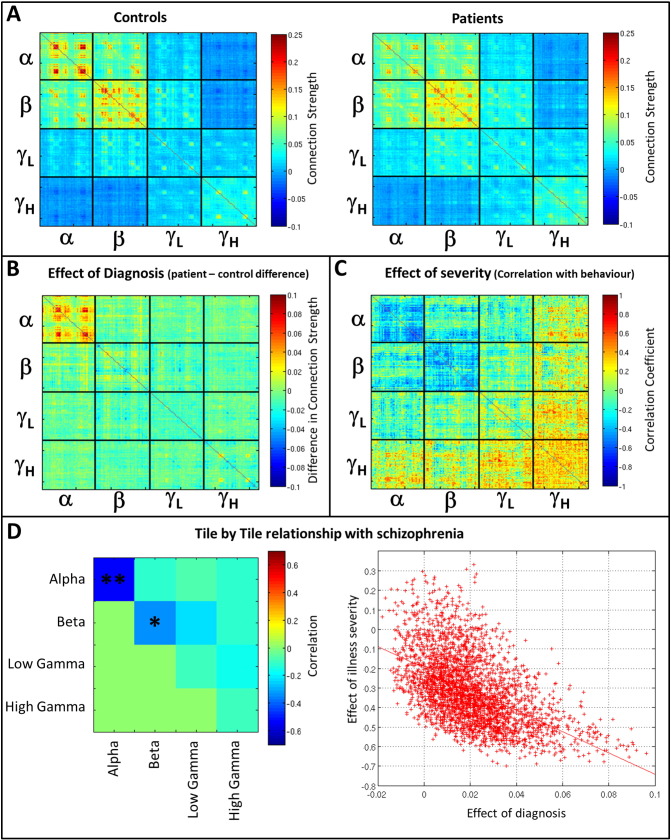
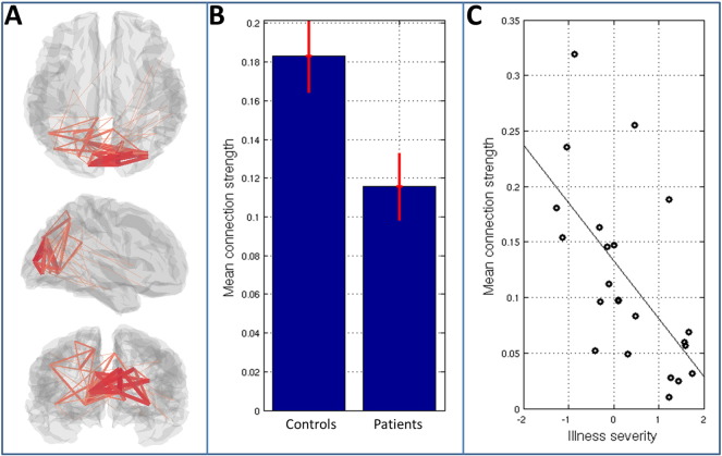
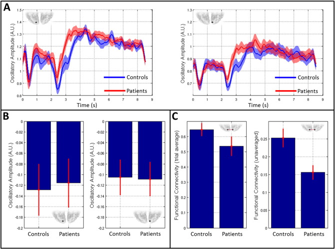
Recent years have shown the critical importance of inter-regional neural network connectivity in supporting healthy brain function. Such connectivity is measurable using neuroimaging techniques such as MEG, however the richness of the electrophysiological signal makes gaining a complete picture challenging. Specifically, connectivity can be calculated as statistical interdependencies between neural oscillations within a large range of different frequency bands. Further, connectivity can be computed between frequency bands. This pan-spectral network hierarchy likely helps to mediate simultaneous formation of multiple brain networks, which support ongoing task demand. However, to date it has been largely overlooked, with many electrophysiological functional connectivity studies treating individual frequency bands in isolation. Here, we combine oscillatory envelope based functional connectivity metrics with a multi-layer network framework in order to derive a more complete picture of connectivity within and between frequencies. We test this methodology using MEG data recorded during a visuomotor task, highlighting simultaneous and transient formation of motor networks in the beta band, visual networks in the gamma band and a beta to gamma interaction. Having tested our method, we use it to demonstrate differences in occipital alpha band connectivity in patients with schizophrenia compared to healthy controls. We further show that these connectivity differences are predictive of the severity of persistent symptoms of the disease, highlighting their clinical relevance. Our findings demonstrate the unique potential of MEG to characterise neural network formation and dissolution. Further, we add weight to the argument that dysconnectivity is a core feature of the neuropathology underlying schizophrenia.

Keywords: Functional connectivity; MEG; Magnetoencephalography; Motor cortex; Multi-layer networks; Neural oscillations; Schizophrenia; Visual cortex.


Figures

Fig. A1
Fig. A1
Testing for leakage and signal to noise ratio between control and patient groups. A) Adjacency matrices representing source leakage between AAL regions. Note that whilst leakage is apparent, there is little difference between groups. B) Summed leakage between all brain regions for controls and patients. C) Regional specific leakage between left and right visual cortex. D) Measured SNR in left visual area. E) Measured SNR in right visual area. Note neither leakage nor SNR differs between groups.
Fig. 1
Fig. 1
Schematic diagram of the connectivity data analysis pipeline including construction of a multi-layer network. Note that, in our actual analysis, the gamma band was split into two, separating low gamma (30 Hz–50 Hz) and high gamma (50–100 Hz). However in order to simplify the Figure, this is not shown.
Fig. 2
Fig. 2
Task induced changes in oscillatory amplitude. A) TFSs generated in the left primary sensorimotor region, and left primary visual area, in healthy control subjects. B) AAL regions exhibiting a significant (pc < 0.05) change in oscillatory amplitude between stimulus and rebound windows. The four images show the four separate frequency bands studied (alpha, beta, low gamma and high gamma).
Fig. 3
Fig. 3
Task induced change in functional connectivity. A) Schematic showing structure of each individual tile (upper panel) and how these are combined to form the super-adjacency matrix (lower panel). B) Super-adjacency matrices computed in the active (left) and control (right) time windows. Matrices show within frequency band (diagonal tiles) and between frequency band (off diagonal tiles) interactions. C) Task induced change (Active–Control) in connectivity. The left hand panel shows change averaged across all subjects. The right hand panel shows the same matrix thresholded to include only statistically significant (pc < 0.01 – FDR corrected) changes in connectivity. Note the main differences occur in the beta and high gamma bands, with significant between frequency interactions in the beta to low gamma, and beta to high gamma bands.
Fig. 4
Fig. 4
Visualisation of task induced change in functional connectivity. The central matrix depicts the dSM, whilst the outer images show significant task induced changes within individual tiles. Significant results are observed in the beta and high gamma bands, as well as between frequency band effects in the beta to low gamma, and beta to high gamma ranges. In all images, the line thickness represents the magnitude of task induced connectivity change.
Fig. 5
Fig. 5
Differences in functional connectivity between controls and schizophrenia patients. A) Super-adjacency matrices computed in controls (left) and patients (right). B) Effect of diagnosis (i.e. difference in connectivity between groups (Controls–Patients)). C) Effect of severity (correlation across individuals between connectivity and the severity of persistent symptoms of schizophrenia, measured by questionnaire). D) Tile correlation showing the relationship between the effects of diagnosis and effects of severity. Relationships are measured as Pearson correlation coefficients across all matrix elements within each tile of the super-adjacency matrix. ** indicates a significant correlation (pc < 0.05 corrected for multiple comparisons across tiles). * indicates a trend (p < 0.05 uncorrected). The right hand panel shows the single example of correlation across matrix elements in the alpha band (p = 0.0005).
Fig. 6
Fig. 6
Visualisation of the differences in alpha band functional connectivity between patients and controls. A) Shows the brain regions between which connectivity differs most between groups. The line width represents the strength of the difference. B) Mean connection strength, averaged across the network identified in (A), for patients and controls. Error bars represent standard error across subjects. C) Mean connection strength (again averaged over all connections in (A)) computed in 23 patients and plotted against a measure of illness severity.
Fig. 7
Fig. 7
Alpha band amplitude and connectivity changes in visual cortex. A) Timecourses of alpha band oscillatory envelope in patients (red) and controls (blue). The left hand plot shows left visual cortex whereas the right hand plot shows right visual cortex. B) The left and right bar charts show mean task induced change in alpha band oscillatory amplitude in left and right visual cortices respectively. C) The left hand plot shows alpha “connectivity” between left and right visual cortices, calculated using trial averaged data (i.e. correlation between the trial averaged alpha envelopes). The right hand bar chart shows alpha connectivity between left and right visual regions based on unaveraged data. Note a significant difference in connectivity when calculated using unaveraged data. This is measured in the absence of measurable differences in task induced amplitude change or a significant change in trial averaged correlation.

References

    1. Adjamian P., Holliday I.E., Barnes G.R., Hillebrand A., Hadjipapas A., Singh K.D. Induced visual illusions and gamma oscillations in human primary visual cortex. Eur. J. Neurosci. 2004;20(2):587–592. - PubMed
    1. Baker A.P., Brookes M.J., Smith S.M., Beherens T., Probert Smith P.J., Woolrich M. Proceedings of the 18th Annual meeting of the organisation for human brain mapping Beijing. 2012. Investigating the temporal dynamics of resting state brain connectivity using magnetoencephalography.
    1. Baker A.P., Brookes M.J., Rezek I.A., Smith S.M., Behrens T., Probert Smith P.J., Woolrich M. Fast transient networks in spontaneous human brain activity. Elife. 2014;3 - PMC - PubMed
    1. Beckmann C.F., De Luca M., Devlin J.T., Smith S.M. Investigations into resting-state connectivity using independent component analysis. Philos. Trans. R. Soc. Lond. 2005;360(1457):1001–1013. - PMC - PubMed
    1. Biswal B., Yetkin F.Z., Haughton V.M., Hyde J.S. Functional connectivity in the motor cortex of resting human brain using echo planar MRI. Magn. Reson. Med. 1995;34:537–541. - PubMed

LinkOut - more resources