. 2016 Mar 22;7(12):14925-39.
doi: 10.18632/oncotarget.7483.

Telomere targeting with a novel G-quadruplex-interactive ligand BRACO-19 induces T-loop disassembly and telomerase displacement in human glioblastoma cells

Affiliations

Telomere targeting with a novel G-quadruplex-interactive ligand BRACO-19 induces T-loop disassembly and telomerase displacement in human glioblastoma cells

Guangtong Zhou et al. Oncotarget. .

Abstract

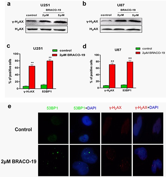
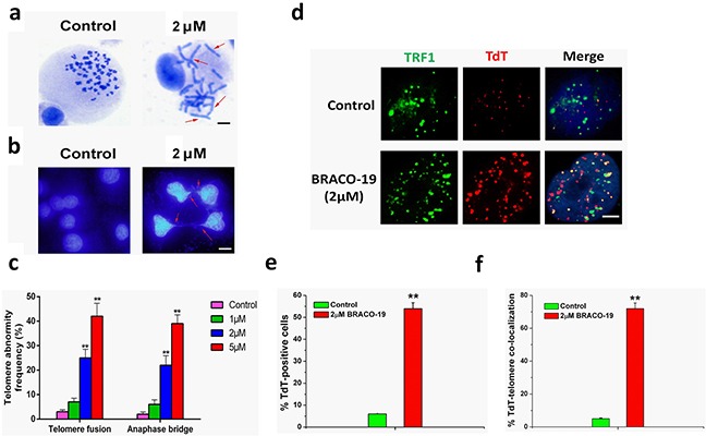
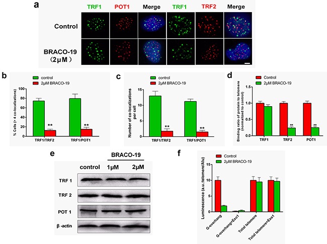
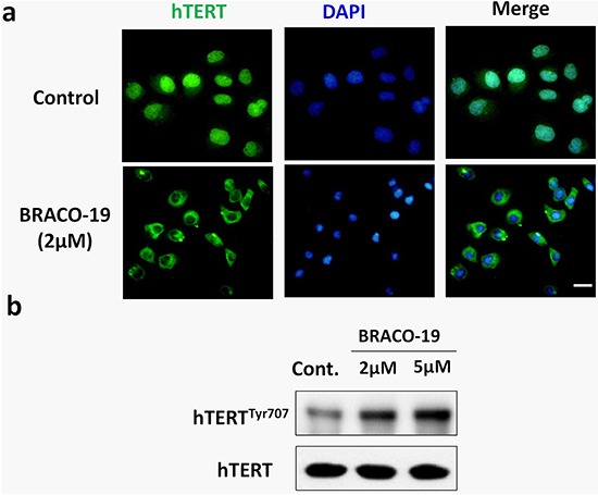
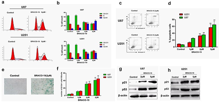
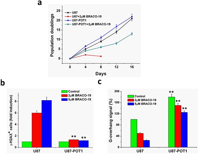
Interference with telomerase and telomere maintenance is emerging as an attractive target for anticancer therapies. Ligand-induced stabilization of G-quadruplex formation by the telomeric DNA 3'-overhang inhibits telomerase from catalyzing telomeric DNA synthesis and from capping telomeric ends, making these ligands good candidates for chemotherapeutic purposes. BRACO-19 is one of the most effective and specific ligand for telomeric G4. It is shown here that BRACO-19 suppresses proliferation and reduces telomerase activity in human glioblastoma cells, paralleled by the displacement of telomerase from nuclear to cytoplasm. Meanwhile, BRACO-19 triggers extensive DNA damage response at telomere, which may result from uncapping and disassembly of telomeric T-loop structure, characterized by the formation of anaphase bridge and telomere fusion, as well as the release of telomere-binding protein from telomere. The resulting dysfunctional telomere ultimately provokes p53 and p21-mediated cell cycle arrest, apoptosis and senescence. Notably, normal primary astrocytes do not respond to the treatment of BRACO-19, suggesting the agent's good selectivity for cancer cells. These results reinforce the notion that G-quadruplex binding compounds can act as broad inhibitors of telomere-related processes and have potential as selective antineoplastic drugs for various tumors including malignant gliomas.

Keywords: DNA damage; G-quadruplex; T-loop; telomerase; telomere.


Conflict of interest statement

CONFLICTS OF INTEREST

The authors declare no potential conflicts of interest.

Figures

Figure 1
Figure 1. Acute BRACO-19 exposure inhibits proliferation and telomerase activity of high grade brain tumor cells
a. U87, U251 and SHG44 cells exhibited IC50 values of 1.45, 1.55 and 2.5 μM respectively when 0.05–25μM BRACO-19 was used, representing a significant inhibition of cell proliferation (P<0.05 for each drug concentration versus untreated). C6 cells exhibited IC50 values of 27.8 μM, showed only a modest cytotoxic effect. b. Telomerase activity inhibition induced by BRACO-19 in U87 and U251 cells. Cells were treated with increasing concentrations of BRACO-19 for 72 hours, CHAPS extract was prepared and equivalent amounts of protein (500 ng) were subjected to a standard TRAP assay. The position of the internal standard was indicated as IS. c. Telomerase activity was quantitated as the percent of the corresponding control sample. The mean of three independent experiments with comparable results was shown. Error bars indicate ± s.d. **P< 0.001, two-tailed student's t-test.
Figure 2
Figure 2. BRACO-19 induces the production of DNA damage response
a, b. Western blot analysis of γ-H2AX in U251 and U87 cells treated with BRACO-19 (2 μM and 5μM) for 72 hours. The levels of H2AX were used as loading control. c, d. Percentage of cells containing γ-H2AX and 53BP1 foci in U251 and U87 cells treated with BRACO-19 (2 μM) for 72 hours. γ-H2AX and 53BP1 foci were quantified using mouse monoclonal antibodies. On average, more than 200 cells were screened in three independent experiments. Error bars indicate s.d. **P< 0.001, two-tailed student's t-test. e. Representative immunofluorescence images of γ-H2AX and 53BP1 foci in U87 cells treated with BRACO-19 (2 μM) for 72 hours. Scale bar equals 5 μm.
Figure 3
Figure 3. DNA-damage response triggered by BRACO-19 occurred at telomeres
a. U87 cells treated with BRACO-19 (2 μM) for 72 hours were fixed and processed for immunofluorescence using antibodies against γ-H2AX (red)/TRF1 (green) or 53BP1 (green)/TRF1 (red), respectively. Representative confocal images were shown. Scale bar equals 5 μm. b. TIF index, defined as foci of DNA-damage response factors that coincided with TRF1, was calculated as the percentage of TIF-positive cells in U87 cells treated with BRACO-19 (2μM). Cells with four or more γ-H2AX/TRF1or 53BP1/TRF1 foci were scored as TIF-positive. Error bars indicate s.d. **P < 0.001, two-tailed student's t-test. c. Average number of TIFs per nucleus in U87 cells treated with BRACO-19 (2μM). Error bars indicated ± s.d. **P < 0.005, two-tailed student's t-test. d. Binding of γ-H2AX and 53BP1 was examined by ChIP assay and detected by qRT–PCR amplification of the telomeric region in U87 cells treated with BRACO-19 (2μM). Data represented triplicate ChIP experiments, each with technical triplicates of qRT–PCR; **P < 0.01 as compared with controls.
Figure 4
Figure 4. Telomere uncapping induced by BRACO-19
a. Telomere fusion induced by BRACO-19. Metaphase spreads were stained with Giemsa. Scale bar equals 10 μm. b. Representative images of anaphase bridges in U87 cells treated with BRACO-19 (2μM) for 72 hours were shown. Cells were stained with DAPI and images were recorded. Red arrow indicated bridge formation. Scale bar equals 10 μm. c. The frequency of telomere instability was calculated as the ratio between cells exhibiting anaphase bridges and the total number of anaphase cells (at least 50 anaphase cells were examined). Telomeric fusion frequency was calculated as total number of telomeric fusions/total number of metaphases. **P < 0.001. d. BRACO-19 induced accessible telomere ends. TRF1 (green) were used to detect telomeres, whereas TdT-cy3 (red) was used as a marker of uncapped telomeres in U87 cells treated with BRACO-19. Merged signals were shown in the right. Scale bar equals 2 μm. e. Quantification of the percentage of TdT-cy3-positive cells in BRACO-19 -treated cells. f. Quantification of the percentage of co-localization of telomeric signals with TdT-cy3 signals in BRACO-treated cells. In panels e and f, a minimum of 100 nuclei was scored, and error bars represented s.d. **P < 0.001.
Figure 5
Figure 5. BRACO-19 specifically delocalizes TRF2 and POT1 from telomeres and induces telomeric 3′-overhang degradation
a. U87 cells treated with BRACO-19 (2 μM) for 72 hours were double stained with the indicated antibodies. Representative confocal images showing merged TRF1 (green) with TRF2 and POT1 (red) staining in untreated and treated cells. Scale bar equals 5 μm. b. Percentages of cells with more than four co-localizations per nucleus of TRF1/TRF2 and TRF1/POT1. Error bars indicated s.d. **P < 0.005. c. Average number of co-localizations per nucleus in U87 cells treated with BRACO-19 (2 μM). Error bars indicated ± s.d. **P < 0.005, two-tailed student's t-test. d. Binding of TRF1, TRF2 or POT1 was examined by ChIP assay and detected by qRT–PCR amplification of the telomeric region in U87 cells treated with BRACO-19 (2 μM). Data represented triplicate ChIP experiments, each with technical triplicates of qRT–PCR; **P < 0.01 as compared with controls. e. Expression of TRF1, TRF2 and POT1 in U87 cells treated with BRACO-19 (2 μM). β-actin was used as loading control. f. Hybridization protection assay (HPA) was performed on genomic DNA isolated from U87 cells treated with BRACO-19 (2 μM) to assess the length of G-overhang and total telomere length. ExoI nuclease digestion was used to assess integrity of the 3′-overhang. Luminescence intensity in arbitrary units (AU) was normalized against Alu probe. Error bars indicated ± s.d., **P < 0.01, two-tailed student's t-test.
Figure 6
Figure 6. BRACO-19 treatment leads to a decrease of hTERT expression in the nucleus and translocation to cytoplasm
a. U87 cells treated with BRACO-19 were stained with the hTERT antibodies for hTERT (green) and DAPI for nucleus (blue). Representative confocal images were shown. Scale bar equals 20 μm. b. Western blot analysis of hTERT phosphorylation in cells exposure to BRACO-19. The levels of hTERT were used as loading control.
Figure 7
Figure 7. Cell cycle arrest, apoptosis and senescence evoked by BRACO-19-induced telomere dysfunction
a, b. Cell cycle arrest induced by BRACO-19 in U87 and U251 cells. 72 hours after treatment with BRACO-19, cells were collected and stained with propidium iodide (PI); DNA content was determined by flow cytometry. **P < 0.01. c, d. Apoptotic cell death induced by BRACO-19 in U87 and U251 cells. 72 hours after treatment, cells were collected and stained with PI and Annexin V–FITC, Annexin V-positive/PI-negative cells were measured by flow cytometry. **P < 0.01. e. Representative images of SA β-gal positive cells in U87 cells that treated with BRACO-19 (2 μM) and control groups. Scale bar equals 50 μm. f. Expression of senescence-associated β-galactosidase (SA-β-gal) in U87 and U251 cells after treatment with BRACO-19 for 72 hours. The senescent cells were counted under an inverted microscope in five random fields. **P < 0.001. g, h. Upregulation of p53 and p21 proteins induced by BRACO-19. Immunoblotting for β-actin was performed to verify equivalent protein loading.
Figure 8
Figure 8. Overexpression of POT1 increases G-overhang and protects cells from BRACO-19 effects
a. Proliferation curves of U87 and U87-POT1 cells treated with BRACO-19 (2μM). At the indicated times, cells were counted and the Population doublings (PDs) were determined. b. POT1 overexpression antagonizes BRACO-19 induced damage response. U87 and U87-POT1 cells were treated with BRACO-19 (2μM, 5μM) for 72h and processed for IF. Histogram represents the increase of γ-H2AX-positive cells compared to the untreated ones. Error bars represent s.d. c. G-overhang signal of U87 cells transfected with POT1 in the absence or treated with BRACO-19 (2 and 5μM) for 72 h. Hybridization protection assay (HPA) was performed on genomic DNA isolated from U87 and U87-POT1 cells to assess the length of G-overhang. Luminescence intensity in arbitrary units (AU) was normalized against Alu probe. Error bars indicated s.d.

References

    1. de Lange T. How telomeres solve the end-protection problem. Science. 2009;326:948–952. - PMC - PubMed
    1. O'Sullivan RJ, Karlseder J. Telomeres: protecting chromosomes against genome instability. Nat Rev Mol Cell Biol. 2010;11:171–181. - PMC - PubMed
    1. Neidle S, Parkinson GN. The structure of telomeric DNA. Curr Opin Struct Biol. 2003;13:275–283. - PubMed
    1. Harley CB, Futcher AB, Greider CW. Telomeres shorten during ageing of human fibroblasts. Nature. 1990;345:458–460. - PubMed
    1. Kim NW, Piatyszek MA, Prowse KR, Harley CB, West MD, Ho PL, Coviello GM, Wright WE, Weinrich SL, Shay JW. Specific association of human telomerase activity with immortal cells and cancer. Science. 1994;266:2011–2015. - PubMed

LinkOut - more resources