. 2016 Feb 23;7(1):e02154-15.
doi: 10.1128/mBio.02154-15.

Host-Primed Ebola Virus GP Exposes a Hydrophobic NPC1 Receptor-Binding Pocket, Revealing a Target for Broadly Neutralizing Antibodies

Affiliations

Host-Primed Ebola Virus GP Exposes a Hydrophobic NPC1 Receptor-Binding Pocket, Revealing a Target for Broadly Neutralizing Antibodies

Zachary A Bornholdt et al. mBio. .

Abstract

The filovirus surface glycoprotein (GP) mediates viral entry into host cells. Following viral internalization into endosomes, GP is cleaved by host cysteine proteases to expose a receptor-binding site (RBS) that is otherwise hidden from immune surveillance. Here, we present the crystal structure of proteolytically cleaved Ebola virus GP to a resolution of 3.3 Å. We use this structure in conjunction with functional analysis of a large panel of pseudotyped viruses bearing mutant GP proteins to map the Ebola virus GP endosomal RBS at molecular resolution. Our studies indicate that binding of GP to its endosomal receptor Niemann-Pick C1 occurs in two distinct stages: the initial electrostatic interactions are followed by specific interactions with a hydrophobic trough that is exposed on the endosomally cleaved GP1 subunit. Finally, we demonstrate that monoclonal antibodies targeting the filovirus RBS neutralize all known filovirus GPs, making this conserved pocket a promising target for the development of panfilovirus therapeutics.

Importance: Ebola virus uses its glycoprotein (GP) to enter new host cells. During entry, GP must be cleaved by human enzymes in order for receptor binding to occur. Here, we provide the crystal structure of the cleaved form of Ebola virus GP. We demonstrate that cleavage exposes a site at the top of GP and that this site binds the critical domain C of the receptor, termed Niemann-Pick C1 (NPC1). We perform mutagenesis to find parts of the site essential for binding NPC1 and map distinct roles for an upper, charged crest and lower, hydrophobic trough in cleaved GP. We find that this 3-dimensional site is conserved across the filovirus family and that antibody directed against this site is able to bind cleaved GP from every filovirus tested and neutralize viruses bearing those GPs.


Figures

FIGURE 1
FIGURE 1
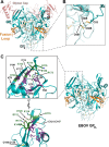
Crystal structure of ebolavirus GPCL. (A) The trimeric EBOV GPCL structure is shown, with GP1 colored teal, GP2 colored light blue, the fusion loop colored orange, and disulfide bonds displayed as sticks and colored gold. The former position of the glycan cap, now absent in the GPCL structure, is illustrated in semitransparent red and is derived from an alignment with the uncleaved EBOV GP structure (PDB code 3CSY). (B) Additional residues at the C terminus of GP2 are now visible in this higher-resolution structure. These residues include C601-C608, contained within GP2, as well as the C53-C609 disulfide bond that cross-links GP1 and GP2 together. (C) The structure of EBOV GPCL is displayed to the right, with the same coloring as described for panel A. An enlarged illustration of the putative EBOV GP1 RBS is shown to the left, in two orientations. Residues forming the hydrophilic crest and hydrophobic trough are labeled and colored green and purple, respectively. The disulfide bonds present around the crest and trough, C108-C135 and C121-C145, are colored gold.
FIGURE 2
FIGURE 2
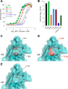
Mutagenic occlusion of the EBOV GP1 receptor-binding site. (A) Alanine or methionine mutations were made to key residues in the RBS. The affinities of wild-type and mutant GPCL for NPC1 domain C were analyzed via ELISA. Note that the L122A and T83M+I113M mutations significantly reduce binding to NPC1 domain C. Means ± SD (n = 4) from a representative experiment are shown. (B) Graph displaying titers of VSV pseudoviruses harboring GP1 RBS mutations. Means ± SD (n = 2–4) from a representative experiment are shown. (C) A semitransparent surface has been placed over the cartoon model of the WT RBS on EBOV GP1 to display the RBS pocket (within the dashed oval outline). Residues T83 and I113 are illustrated as sticks (black). (D) Model of EBOV RBS bearing the mutations T83M and I113M (red). The longer side chains of the introduced methionine residues fill the RBS pocket and likely prevent NPC1 domain C binding by occluding the NPC1 binding site. (E) The buried location of L122 (black) is displayed in the EBOV GP1 RBS. See also Fig. S1, S2, and S4 in the supplemental material.
FIGURE 3
FIGURE 3
The basic electrostatic potential of the GP1 crest is vital to receptor binding. (A) ELISA analysis of binding of wild-type or mutant GPCL to NPC1 domain C. Replacement of positively charged K114 and K115 with neutral alanines or negatively charged glutamic acids reduces and abrogates NPC1 binding, respectively. Concomitant mutation of neighboring E112 and E120 to neutral alanine residues restores affinity for NPC1 domain C. Means ± SD (n = 4) from a representative experiment are shown. (B) Growth titers of VSV pseudotyped with electrostatic mutants of EBOV GP correlate with the NPC1 domain C affinities shown by the results in panel A: reduction in growth correlates with loss of positive charge. Means ± SD (n = 2–4) from a representative experiment are shown. (C) The electrostatic surface potential is calculated for each of the mutant EBOV GPs using APBS in PyMol (The PyMOL Molecular Graphics System, version 1.5.0.4. [Schrödinger, LLC], and APBS plugin for PyMol, M. G. Lerner and H. A. Carlson, University of Michigan, Ann Arbor, MI, 2006; 48). The view is looking down onto the EBOV GP trimer. Mutants with an overall negative charge on the surface of GP1 demonstrate defects in both affinity for NPC1 domain C and viral growth. See also Fig. S1, S2, and S4 in the supplemental material.
FIGURE 4
FIGURE 4
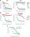
Monoclonal antibodies targeting the conserved GP1 RBS demonstrate panfilovirus neutralization activity. (A) VSV pseudotyped with GPs from different species of filovirus (as indicated in the key to the right) were preprimed with thermolysin to expose the GP1 RBS and then analyzed for reduction in relative infectivity following treatment with MR72 or MR78. (B) The graph to the left shows a comparative analysis of the neutralization of VSV-EBOV GPCL and VSV-EBOV GPCL-V79A by MR72 and MR78. The graph to the right displays the results of competitive binding assays detecting NPC1 domain C binding in the presence of increasing concentrations of MR72 or MR78 for EBOV GPCL and EBOV GPCL-V79A. The key for both graphs is on the far right. (C) Graph showing the results of comparative infectivity assays of nonprimed VSV pseudotyped with EBOV GP treated with MAbs from the ZMapp cocktail (2G4, 4G7, and 13C6) (33) or the neutralizing EBOV antibody KZ52 (21). MR72 neutralizes primed EBOV GPCL pseudovirions at >10-fold lower concentrations than are required for ZMapp or KZ52 to neutralize EBOV GP pseudovirions. See also Fig. S3 in the supplemental material. Means ± SD (n = 2−4) from a representative experiment are shown in each panel.

References

    1. De la Vega MA, Stein D, Kobinger GP. 2015. Ebolavirus evolution: past and present. PLoS Pathog 11:e1005221. doi:10.1371/journal.ppat.1005221. - DOI - PMC - PubMed
    1. Negredo A, Palacios G, Vázquez-Morón S, González F, Dopazo H, Molero F, Juste J, Quetglas J, Savji N, de la Cruz Martínez M, Herrera JE, Pizarro M, Hutchison SK, Echevarría JE, Lipkin WI, Tenorio A. 2011. Discovery of an ebolavirus-like filovirus in Europe. PLoS Pathog 7:e1002304. doi:10.1371/journal.ppat.1002304. - DOI - PMC - PubMed
    1. Baize S, Pannetier D, Oestereich L, Rieger T, Koivogui L, Magassouba N, Soropogui B, Sow MS, Keïta S, De Clerck H, Tiffany A, Dominguez G, Loua M, Traoré A, Kolié M, Malano ER, Heleze E, Bocquin A, Mély S, Raoul H, Caro V, Cadar D, Gabriel M, Pahlmann M, Tappe D, Schmidt-Chanasit J, Impouma B, Diallo AK, Formenty P, Van Herp M, Gunther S. 2014. Emergence of Zaire Ebola virus disease in Guinea. N Engl J Med 371:1418–1425. doi:10.1056/NEJMoa1404505. - DOI - PubMed
    1. Lee JE, Fusco ML, Hessell AJ, Oswald WB, Burton DR, Saphire EO. 2008. Structure of the Ebola virus glycoprotein bound to an antibody from a human survivor. Nature 454:177–182. doi:10.1038/nature07082. - DOI - PMC - PubMed
    1. Lee JE, Saphire EO. 2009. Neutralizing ebolavirus: structural insights into the envelope glycoprotein and antibodies targeted against it. Curr Opin Struct Biol 19:408–417. doi:10.1016/j.sbi.2009.05.004. - DOI - PMC - PubMed

Publication types

MeSH terms

Associated data