. 2016 Apr 15;25(8):1479-88.
doi: 10.1093/hmg/ddw022. Epub 2016 Jan 28.

Mutations in POMGNT1 cause non-syndromic retinitis pigmentosa

Affiliations

Mutations in POMGNT1 cause non-syndromic retinitis pigmentosa

Mingchu Xu et al. Hum Mol Genet. .

Abstract

A growing number of human diseases have been linked to defects in protein glycosylation that affects a wide range of organs. Among them, O-mannosylation is an unusual type of protein glycosylation that is largely restricted to the muscular and nerve system. Consistently, mutations in genes involved in the O-mannosylation pathway result in infantile-onset, severe developmental defects involving skeleton muscle, brain and eye, such as the muscle-eye-brain disease (MIM no. 253280). However, the functional importance of O-mannosylation in these tissues at later stages remains largely unknown. In our study, we have identified recessive mutations in POMGNT1, which encodes an essential component in O-mannosylation pathway, in three unrelated families with autosomal recessive retinitis pigmentosa (RP), but without extraocular involvement. Enzymatic assay of these mutant alleles demonstrate that they greatly reduce the POMGNT1 enzymatic activity and are likely to be hypomorphic. Immunohistochemistry shows that POMGNT1 is specifically expressed in photoreceptor basal body. Taken together, our work identifies a novel disease-causing gene for RP and indicates that proper protein O-mannosylation is not only essential for early organ development, but also important for maintaining survival and function of the highly specialized retinal cells at later stages.


Figures

Figure 1.
Figure 1.
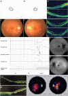
Clinical findings of non-syndromic RP patients in this study. Visual field test results of MOGL2064, right eye (A) and left eye (B), showing restricted visual field in both eyes. OCT images of MOGL2064, right eye (C) and left eye (D), featuring chorioretinal atrophy, retinal thinning with absent IS/OS junctions. Fundus images of SRF1105, right eye (E) and left eye (F), showing the tigroid appearance, thinning of mid-peripheral RPE and loss of choroid-capillaries. OCT images of SRF1105, right eye (G) and left eye (H), showing thinning of outer retinal layer with preserved foveal photoreceptors. (I) ERG results of SRF1105, showing no detectable ERG signals. Fluorescein angiography results of SRF1105, right eye (J) and left eye (K), showing oval shaped hypofluorescence with a hyperfluorescent ring in the macular and mottled hypofluorescence area in the peripheral retina. OCT images of SRF1630, right eye (L) and left eye (M), showing thinning of whole retina layers, a flat fovea and disappearance of IS/OS (right eye more severe than left eye). Visual field test results of patient SRF1630, right eye (N) and left eye (O), showing tunnel vision in both eyes.
Figure 2.
Figure 2.
Genetic findings of non-syndromic RP patients in this study. (A) Four non-syndromic RP patients from three unrelated families with POMGNT1 biallelic variants were identified. Genotypes (cDNA and protein changes based on reference cDNA sequence NM_017739) are labeled for each family. (B) POMGNT1 protein sequence alignment among selected vertebrates at the loci of POMGNT1 missense variants identified in our study. All the disrupted amino acids are highly conserved in vertebrates.
Figure 3.
Figure 3.
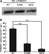
Biochemical studies to test the pathogenicity of two POMGNT1 missense variants. (A) Western blot analysis of POMGNT1 WT and mutant proteins, showing their expression and approximately equal molecular weights. (B) Enzymatic activity of POMGNT1 WT and mutant proteins. Both mutants retained significantly lower activity compared with the WT level. Two-tailed Welch's t-test. ***P < 10−5. Error bars indicate sample standard deviations.
Figure 4.
Figure 4.
POMGNT1 is expressed in photoreceptor cells. For each individual photoreceptor, the POMGNT1 signal is next to the cilia marker (acetylated-α-tubulin) signal. Two-dot pattern (labeled by double arrows) indicates the localization of POMGNT1 at both the basal body and the daughter centriole of photoreceptor cells. Ac-α-Tub, acetylated-α-tubulin; OS, outer segments; CC, connecting cilium; IS, inner segments; ONL, outer nuclear layer.

References

    1. Ferrari S., Di Iorio E., Barbaro V., Ponzin D., Sorrentino F.S., Parmeggiani F. (2011) Retinitis pigmentosa: genes and disease mechanisms. Curr. Genomics, 12, 238–249. - PMC - PubMed
    1. Wang F., Wang H., Tuan H.F., Nguyen D.H., Sun V., Keser V., Bowne S.J., Sullivan L.S., Luo H., Zhao L. et al. (2014) Next generation sequencing-based molecular diagnosis of retinitis pigmentosa: identification of a novel genotype-phenotype correlation and clinical refinements. Hum. Genet., 133, 331–345. - PMC - PubMed
    1. Rachel R.A., Li T., Swaroop A. (2012) Photoreceptor sensory cilia and ciliopathies: focus on CEP290, RPGR and their interacting proteins. Cilia, 1, 22. - PMC - PubMed
    1. Haltia M. (2006) The neuronal ceroid-lipofuscinoses: from past to present. Biochim. Biophys. Acta, 1762, 850–856. - PubMed
    1. Hayflick S.J. (2003) Unraveling the Hallervorden-Spatz syndrome: pantothenate kinase-associated neurodegeneration is the name. Curr. Opin. Pediatr., 15, 572–577. - PubMed

Publication types

MeSH terms

Substances