. 2016 Apr 1;25(7):1370-81.
doi: 10.1093/hmg/ddw019. Epub 2016 Jan 28.

A critical period of vulnerability to adolescent stress: epigenetic mediators in mesocortical dopaminergic neurons

Affiliations

A critical period of vulnerability to adolescent stress: epigenetic mediators in mesocortical dopaminergic neurons

Minae Niwa et al. Hum Mol Genet. .

Abstract

The molecular basis of vulnerability to stress during the adolescent period is largely unknown. To identify potential molecular mediators that may play a role in stress-induced behavioral deficits, we imposed social isolation on a genetically vulnerable mouse model. We report that 3-week (5-8 weeks of age) adolescent stress in combination with disrupted-in-schizophrenia 1 (Disc1) genetic risk elicits alterations in DNA methylation of a specific set of genes, tyrosine hydroxylase, brain-derived neurotrophic factor and FK506 binding protein 5. The epigenetic changes in the mesocortical dopaminergic neurons were prevented when animals were treated with a glucocorticoid receptor (GR) antagonist RU486 during social isolation, which implicates the role for glucocorticoid signaling in this pathological event. We define the critical period of GR intervention as the first 1-week period during the stress regimen, suggesting that this particular week in adolescence may be a specific period of maturation and function of mesocortical dopaminergic neurons and their sensitivity to glucocorticoids. Our study may also imply the clinical significance of early detection and prophylactic intervention against conditions associated with adolescent social stress in individuals with genetic risk.


Figures

Figure 1.
Figure 1.
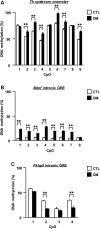
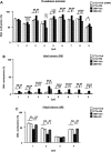
Influence of adolescent isolation on epigenetic modifications of the Th, Bdnf and Fkbp5 genes in VTA neurons that project to the frontal cortex between CTL and DM. (A) DNA methylation in the promoter of Th. (B) DNA methylation in the intronic GRE of Bdnf. (C) DNA methylation in the intronic GRE of Fkbp5. CTL, wild-type mice without isolation. DM, DISC1 mutant exposed to 3-week isolation. N = 3. Each sample was pooled from five males. Values are means ± SEM. Statistical differences were detected using two-way ANOVA with repeated measures (group, F(1,4) = 52.34, P< 0.01 for A; F(1,4) = 444.3, P< 0.01 for B; F(1,4) = 53.24, P< 0.01 for C), followed by Bonferroni post hoc tests (**P< 0.01).
Figure 2.
Figure 2.
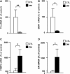
Influence of adolescent isolation on mRNA expression of Th, Bdnf, Fkbp5 and GR genes in VTA neurons that project to the frontal cortex between CTL and DM. (A) Expression levels of Th mRNA. (B) Expression levels of Bdnf mRNA. (C) Expression levels of Fkbp5 mRNA. (D) Expression levels of Gr mRNA. Expression levels of β-Actin were used as an internal control. N = 4–8. Each sample was pooled from five males. Values are means ± SEM. Statistical differences between two groups were analyzed with the t-test (**P< 0.01 and *P< 0.05).
Figure 3.
Figure 3.
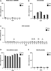
No influence of adolescent isolation on genes involved in the HPA-axis and other signaling pathways in VTA neurons that project to the frontal cortex between CTL and DM. (A) DNA methylation in the intronic GRE/ERE of Fkbp4. (B) DNA methylation in the promoter of Crh. (C) DNA methylation in the promoter of Crhr1. (D) DNA methylation in the upstream region of Lepr. (E) DNA methylation in the promoter of Gsk3β. N = 3. Each sample was pooled from five males. Values are means ± SEM. No statistical differences were detected using two-way ANOVA with repeated measures (group, F(1,4) = 0.04, P = 0.86 for A; F(1,4) = 1.21, P = 0.33 for B; F(1,4) = 44.73, P < 0.01 for C; F(1,4) = 0.69, P = 0.45 for D; F(1,4) = 0.00, P = 0.96 for E).
Figure 4.
Figure 4.
Influence of glucocorticoids from 5 to 8 weeks of age on epigenetic modifications of the Th, Bdnf and Fkbp5 genes in the DM. (A) Effects of RU486 on increased DNA methylation in the promoter of Th. (B) Effects of RU486 on increased DNA methylation in the intronic GRE of Bdnf. (C) Effects of RU486 on decreased DNA methylation in the intronic GRE of Fkbp5. N = 3. Each sample was pooled from five males. Values are means ± SEM. Statistical differences were detected using three-way ANOVA with repeated measures (group × drug, F(1,8) = 1798.03, P< 0.01 for A; F(1,8) = 730.52, P< 0.01 for B; F(1,8) = 149.29, P< 0.01 for C) and two-way ANOVA (group × drug, F(1,8) = 0.68, P = 0.43 for CpG1 in A; F(1,8) = 685.10, P< 0.01 for CpG2 in A; F(1,8) = 183.80, P < 0.01 for CpG3 in A; F(1,8) = 125.80, P< 0.01 for CpG4 in A; F(1,8) = 15.55, P< 0.01 for CpG5 in A; F(1,8) = 104.00, P< 0.01 for CpG6 in A; F(1,8) = 1307.00, P< 0.01 for CpG7 in A; F(1,8) = 75.72, P< 0.01 for CpG8 in A; F(1,8) = 154.60, P< 0.01 for CpG9 in A; F(1,8) = 89.96, P< 0.01 for CpG1 in B; F(1,8) = 54.55, P< 0.01 for CpG2 in B; F(1,8) = 43.26, P< 0.01 for CpG3 in B; F(1,8) = 5.73, P< 0.05 for CpG4 in B; F(1,8) = 7.60, P< 0.05 for CpG5 in B; F(1,8) = 89.89, P< 0.01 for CpG6 in B; F(1,8) = 85.22, P< 0.01 for CpG7 in B; F(1,8) = 21.23, P< 0.01 for CpG8 in B; F(1,8) = 12.30, P< 0.01 for CpG1 in C; F(1,8) = 306.60, P< 0.01 for CpG2 in C; F(1,8) = 2.77, P = 0.13 for CpG3 in C; F(1,8) = 44.16, P< 0.01 for CpG4 in C), followed by Bonferroni post hoc tests (**P< 0.01 and *P< 0.05).
Figure 5.
Figure 5.
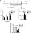
Influence of glucocorticoids from 5 to 7 weeks on behavioral and neurochemical abnormalities in the DM. (A) CTL and DM were treated with RU486 from 5 to 7 weeks of age. (B) Effects of RU486 on performance of prepulse inhibition. (C) Effects of RU486 on performance of the forced swim test. (D) Effects of RU486 on basal levels of extracellular dopamine in the frontal cortex. Veh, treated with vehicle; RU, treated with RU486. N = 11–13 for (B and C), N = 6 for (D). Values are means ± SEM. Statistical differences were determined using three-way ANOVA with repeated measures (group × drug, F(1,43) = 3.29, P= 0.08 for B), and two-way ANOVA (group × drug, F(1,43) = 0.42, P = 0.52 for prepulse 74 in B; F(1,43) = 2.19, P= 0.15 for prepulse 78 in B; F(1,43) = 4.08, P< 0.05 for prepulse 86 in B; F(1,43) = 8.37, P< 0.01 for C; F(1,20) = 5.88, P< 0.05 for D), followed by Bonferroni post hoc tests (**P< 0.01 and *P< 0.05).
Figure 6.
Figure 6.
Influence of glucocorticoids from 5 to 6 weeks on behavioral and neurochemical abnormalities in the DM. (A) CTL and DM were treated with RU486 from 5 to 6 weeks of age. (B) Effects of RU486 on performance of prepulse inhibition. (C) Effects of RU486 on performance of the forced swim test. (D) Effects of RU486 on basal levels of extracellular dopamine in the frontal cortex. N = 11 for (B and C), N = 6–10 for (D). Values are means ± SEM. Statistical differences were determined using three-way ANOVA with repeated measures (group × drug, F(1,40) = 2.80, P= 0.10 for B), and two-way ANOVA (group × drug, F(1,40) = 0.48, P = 0.49 for prepulse 74 in B; F(1,40) = 0.91, P= 0.35 for prepulse 78 in B; F(1,40) = 4.25, P< 0.05 for prepulse 86 in B; F(1,40) = 6.70, P< 0.05 for C; F(1,24) = 6.14, P< 0.05 for D), followed by Bonferroni post hoc tests (**P< 0.01 and *P< 0.05).
Figure 7.
Figure 7.
No influence of glucocorticoids from 8 to 10 weeks on behavioral and neurochemical abnormalities in the DM. (A) CTL and DM were treated with RU486 from 8 to 10 weeks of age. (B) Effects of RU486 on performance of prepulse inhibition. (C) Effects of RU486 on performance of the forced swim test. (D) Effects of RU486 on basal levels of extracellular dopamine in the frontal cortex. N = 12–14 for (B and C), N = 6 for (D). Values are means ± SEM. Statistical differences were determined using three-way ANOVA with repeated measures (group × drug, F(1,48) = 0.56, P= 0.46 for B), and two-way ANOVA (group × drug, F(1,48) = 0.00, P = 0.99 for prepulse 74 in B; F(1,48) = 0.82, P= 0.37 for prepulse 78 in B; F(1,48) = 0.44, P= 0.51 for prepulse 86 in B; F(1,48) = 2.06, P= 0.16 for C; F(1,20) = 0.81, P= 0.38 for D).
Figure 8.
Figure 8.
Influence of glucocorticoids from 5 to 6 weeks of age on epigenetic modifications of the Th, Bdnf and Fkbp5 genes in the DM. (A) Effects of RU486 on increased DNA methylation in the promoter of Th. (B) Effects of RU486 on increased DNA methylation in the intronic GRE of Bdnf. (C) Effects of RU486 on decreased DNA methylation in the intronic GRE of Fkbp5. N = 3. Each sample was pooled from five males. Values are means ± SEM. Statistical differences were detected using three-way ANOVA with repeated measures (group × drug, F(1,8) = 358.35, P< 0.01 for A; F(1,8) = 106.99, P< 0.01 for B; F(1,8) = 374.77, P< 0.01 for C), and two-way ANOVA (group × drug, F(1,8) = 0.10, P = 0.76 for CpG1 in A; F(1,8) = 6.61, P< 0.05 for CpG2 in A; F(1,8) = 157.80, P< 0.01 for CpG3 in A; F(1,8) = 17.29, P< 0.01 for CpG4 in A; F(1,8) = 21.96, P< 0.01 for CpG5 in A; F(1,8) = 61.17, P< 0.01 for CpG6 in A; F(1,8) = 67.61, P< 0.01 for CpG7 in A; F(1,8) = 53.16, P< 0.01 for CpG8 in A; F(1,8) = 8.15, P< 0.05 for CpG9 in A; F(1,8) = 21.69, P< 0.01 for CpG1 in B; F(1,8) = 15.53, P< 0.01 for CpG2 in B; F(1,8) = 91.51, P< 0.01 for CpG3 in B; F(1,8) = 6.69, P< 0.05 for CpG4 in B; F(1,8) = 19.85, P< 0.01 for CpG5 in B; F(1,8) = 106.90, P< 0.01 for CpG6 in B; F(1,8) = 56.31, P< 0.01 for CpG7 in B; F(1,8) = 22.46, P< 0.01 for CpG8 in B; F(1,8) = 82.82, P< 0.01 for CpG1 in C; F(1,8) = 83.44, P< 0.01 for CpG2 in C; F(1,8) = 4.40, P = 0.07 for CpG3 in C; F(1,8) = 77.72, P< 0.01 for CpG4 in C), followed by Bonferroni post hoc tests (**P< 0.01 and *P< 0.05).

Similar articles

Cited by

References

    1. Blakemore S.J. (2008) The social brain in adolescence. Nat. Rev. Neurosci., 9, 267–277. - PubMed
    1. Caspi A., Roberts B.W., Shiner R.L. (2005) Personality development: stability and change. Ann. Rev. Psychol., 56, 453–484. - PubMed
    1. Gunnar M., Quevedo K. (2007) The neurobiology of stress and development. Ann. Rev. Psychol., 58, 145–173. - PubMed
    1. Paus T., Keshavan M., Giedd J.N. (2008) Why do many psychiatric disorders emerge during adolescence? Nat. Rev. Neurosci., 9, 947–957. - PMC - PubMed
    1. Niwa M., Jaaro-Peled H., Tankou S., Seshadri S., Hikida T., Matsumoto Y., Cascella N.G., Kano S., Ozaki N., Nabeshima T. et al. (2013) Adolescent stress-induced epigenetic control of dopaminergic neurons via glucocorticoids. Science, 339, 335–339. - PMC - PubMed

Publication types

Substances