. 2016 Mar 22;113(12):3239-44.
doi: 10.1073/pnas.1522236113. Epub 2016 Feb 23.

Phosphorylation and calcium antagonistically tune myosin-binding protein C's structure and function

Affiliations

Phosphorylation and calcium antagonistically tune myosin-binding protein C's structure and function

Michael J Previs et al. Proc Natl Acad Sci U S A. .

Abstract

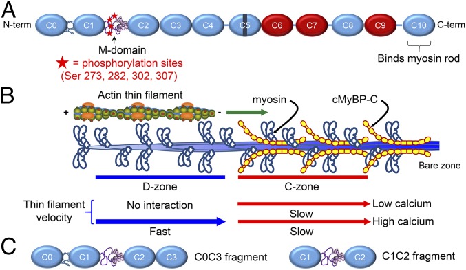
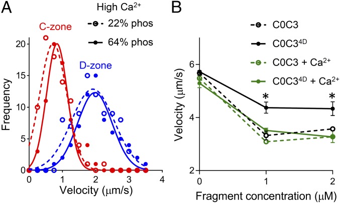
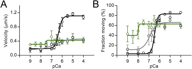
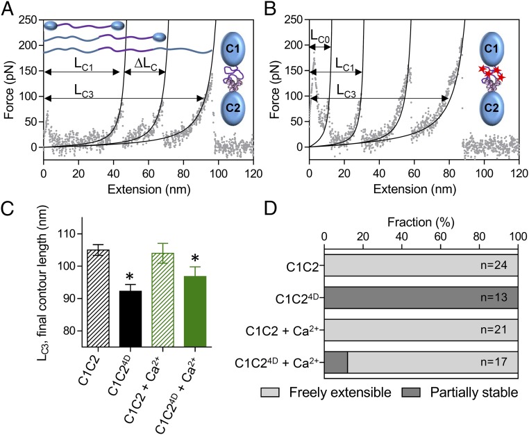
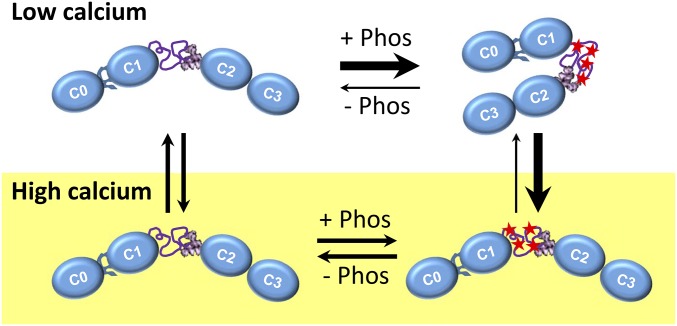
During each heartbeat, cardiac contractility results from calcium-activated sliding of actin thin filaments toward the centers of myosin thick filaments to shorten cellular length. Cardiac myosin-binding protein C (cMyBP-C) is a component of the thick filament that appears to tune these mechanochemical interactions by its N-terminal domains transiently interacting with actin and/or the myosin S2 domain, sensitizing thin filaments to calcium and governing maximal sliding velocity. Both functional mechanisms are potentially further tunable by phosphorylation of an intrinsically disordered, extensible region of cMyBP-C's N terminus, the M-domain. Using atomic force spectroscopy, electron microscopy, and mutant protein expression, we demonstrate that phosphorylation reduced the M-domain's extensibility and shifted the conformation of the N-terminal domain from an extended structure to a compact configuration. In combination with motility assay data, these structural effects of M-domain phosphorylation suggest a mechanism for diminishing the functional potency of individual cMyBP-C molecules. Interestingly, we found that calcium levels necessary to maximally activate the thin filament mitigated the structural effects of phosphorylation by increasing M-domain extensibility and shifting the phosphorylated N-terminal fragments back to the extended state, as if unphosphorylated. Functionally, the addition of calcium to the motility assays ablated the impact of phosphorylation on maximal sliding velocities, fully restoring cMyBP-C's inhibitory capacity. We conclude that M-domain phosphorylation may have its greatest effect on tuning cMyBP-C's calcium-sensitization of thin filaments at the low calcium levels between contractions. Importantly, calcium levels at the peak of contraction would allow cMyBP-C to remain a potent contractile modulator, regardless of cMyBP-C's phosphorylation state.

Keywords: cMyBP-C; muscle activation; muscle regulation; structure-function.


Conflict of interest statement

The authors declare no conflict of interest.

Figures

Fig. 1.
Fig. 1.
Structure and function of cMyBP-C. (A) Schematic diagram of full-length cMyBP-C. Domains C1 and C2 are connected by the M-domain, containing an intrinsically disordered N-terminal region with four phosphorylatable serines, and a more structured C-terminal half. Ig-like domains are shown in blue, and Fn-like domains are shown in red. (B) Illustration of half of a native thick filament with a native thin filament landing on the tip of the thick filament and being translocated through the D- and C-zones at the different speeds indicated, as observed in the TIRFM experiments. (C) Wild-type C0C3 and C1C2 N-terminal fragments used in motility, AFM, and EM assays. Phosphomimetic counterparts to each fragment were expressed containing aspartic acid substitutions for the four serines highlighted in A.
Fig. 2.
Fig. 2.
Effects of cMyBP-C phosphorylation and calcium on actin-filament sliding. (A) Frequency–velocity histograms and Gaussian fits for initial native thin-filament sliding velocity in the D-zone (blue) and C-zone (red) (compare with Fig. 1B). Solid symbols and lines indicate motion on wild-type thick filaments containing highly phosphorylated (∼64%) MyBP-C; open symbols and dashed lines represent motion on thick filaments containing dephosphorylated (∼22%) MyBP-C. Assays were carried out at 100 µM ATP and 22 °C in the presence of 0.01 mM free calcium. (B) Sliding velocities of bare actin filaments on the surface of randomly oriented myosin molecules (skeletal muscle proteins) in the presence of unphosphorylated wild-type C0C3 (open circles, dashed lines) and phosphomimetic C0C34D (closed circles, solid lines), with (green) and without (black) 0.1 mM free calcium. The assay was carried out at 1 mM ATP and 30 °C. *P < 0.01 compared with C0C3, Student’s t test. The impact of cMyBP-C phosphorylation on sliding velocity and its partial reversal by calcium is similar for actomyosin from cardiac (A and Fig. S1) and skeletal (B) muscles, suggesting that calcium acts directly on cMyBP-C.
Fig. S1.
Fig. S1.
The effects of wild-type C0C3 and phosphomimetic C0C34D N-terminal fragments and calcium on mouse cardiac native thin filaments sliding over a surface of randomly oriented mouse cardiac myosin molecules. (A) Velocity and (B) fraction moving versus pCa (−log [Ca2+]) plots (100 µM ATP, 22 °C) for native thin filaments in the absence of cMyBP-C (black symbols and lines) and in the presence of 1 µM wild-type C0C3 (green symbols and lines) and C0C34D (gray symbols and lines). Data were fitted with sigmoidal dose–response curves with variable slopes. Results show that phosphomimetic substitution allows tuning of the activating potential of the C0C3 fragment but has no effect on the modulatory mechanism when the thin filaments are fully activated by calcium.
Fig. 3.
Fig. 3.
Effects of phosphorylation and calcium on the structure of the M-domain. (A and B) AFM force:extension curves for single unphosphorylated wild-type C1C2 (A) and phosphomimetic C1C24D (B) molecules in the absence of calcium. The initial contour length of the freely extensible components (LC1) and final contour length of the fully extended molecules (LC3) are indicated, as is the change in contour length associated with the preceding unfolding event (ΔLC). (C) The final contour lengths of the C1C2 and C1C24D fragments (LC3 in A and B) in the absence and presence 0.1 mM free calcium. *P < 0.01, LC3 vs. C1C2, Student’s t test. (D) The relative stability of the M-domain within the single C1C2 and C1C24D molecules in the absence and presence of 0.1 mM free calcium as determined from the number of molecules showing three (freely extensible M-domain) or four (partially stable M-domain) peaks in the force:extension curves.
Fig. 4.
Fig. 4.
Localization of a hinge point within the M-domain. (A) Negatively stained EM image of full-length baculovirus-expressed cMyBP-C. (Insets) Single molecules with visible Ig and Fn domains. (B) Rotary-shadowed EM image of full-length cMyBP-C molecules. (Insets) Examples of bent rods with one and two hinge points. (C) Rotary-shadowed EM image of full-length cMyBP-C after treatment with calpain to remove the N-terminal domains. (Insets) Molecules with only a single hinge, resulting from site-specific truncation of the N-terminal segment. (D) Schematic diagram of cMyBP-C showing the location of two hinge points connecting more rigid segments.
Fig. S2.
Fig. S2.
Calpain cleavage of full-length cMyBP-C. (A) SDS/PAGE shows that at low calpain concentrations cMyBP-C is cleaved into two major fragments with apparent molecular masses of ∼110 and 40 kDa [confirmed to be 29 kDa by mass spectrometry (1)]. (B) Frequency–length histograms for cMyBP-C segments in elongated wild-type and calpain-cleaved (25:1 ratio) expressed cMyBP-C molecules. Segment lengths for 60 molecules with two hinges and 40 molecules with three hinges were measured for the wild-type molecules (black bars). Segment lengths for 94 calpain-treated cMyBP-C molecules with two hinges and six molecules with three hinges were measured (gray bars). Calpain-treated molecules showed a large reduction in the number with segments ≤8 nm long (segment 1 in Table 1), and only 6% of the calpain-treated molecules showed a terminal bend (three hinges), as compared with 40% of the undigested molecules.
Fig. 5.
Fig. 5.
Effects of phosphorylation and calcium on the orientation of the N-terminal domains. (A and B) Rotary-shadowed EM images of nonphosphorylated wild-type C0C3 (A) and phosphomimetic C0C34D (B) fragments in the absence of calcium. (C) Representative EM images (Left) and schematic diagrams (Right) of the extended rods (Top), bent rods (Middle), and compact/closed structures (Bottom) classified in each EM image. (D and E) Rotary-shadowed EM images as in A and B for C0C3 (D) and C0C34D (E) fragments in the presence of calcium. Yellow, blue, and red arrows point to molecules with extended, bent, and compact structures, respectively.
Fig. S3.
Fig. S3.
The effects of phosphorylation and calcium on the orientation of the N-terminal domains. (A and B) Rotary-shadowed EM images of PKA-treated C0C3 fragments in the absence (A) and presence (B) of 0.1 mM free Ca2+. (C) Blots from a Pro-Q–stained SDS/PAGE gel confirming the presence of phosphate in the PKA-treated sample. As observed for the phosphomimetic C0C34D fragments (Fig. 5 B and E), PKA treatment resulted in molecules adopting a compact conformation, and the presence of calcium shifted these molecules into bent or extended conformations.
Fig. 6.
Fig. 6.
Diagrams of the effects of phosphorylation on the overall structure of cMyBP-C’s N terminus at low (Top) and high (Bottom) calcium levels.

Comment in

References

    1. Harris SP, Lyons RG, Bezold KL. In the thick of it: HCM-causing mutations in myosin binding proteins of the thick filament. Circ Res. 2011;108(6):751–764. - PMC - PubMed
    1. Sequeira V, Witjas-Paalberends ER, Kuster DW, van der Velden J. Cardiac myosin-binding protein C: Hypertrophic cardiomyopathy mutations and structure-function relationships. Pflugers Arch. 2014;466(2):201–206. - PubMed
    1. Sadayappan S, de Tombe PP. Cardiac myosin binding protein-C as a central target of cardiac sarcomere signaling: A special mini review series. Pflugers Arch. 2014;466(2):195–200. - PMC - PubMed
    1. Flashman E, Redwood C, Moolman-Smook J, Watkins H. Cardiac myosin binding protein C: Its role in physiology and disease. Circ Res. 2004;94(10):1279–1289. - PubMed
    1. Craig R, Lee KH, Mun JY, Torre I, Luther PK. Structure, sarcomeric organization, and thin filament binding of cardiac myosin-binding protein-C. Pflugers Arch. 2014;466(3):425–431. - PMC - PubMed

Publication types

LinkOut - more resources