. 2016 Feb 16:10:5.
doi: 10.3389/fncir.2016.00005. eCollection 2016.

A Flight Sensory-Motor to Olfactory Processing Circuit in the Moth Manduca sexta

Affiliations

A Flight Sensory-Motor to Olfactory Processing Circuit in the Moth Manduca sexta

Samual P Bradley et al. Front Neural Circuits. .

Abstract

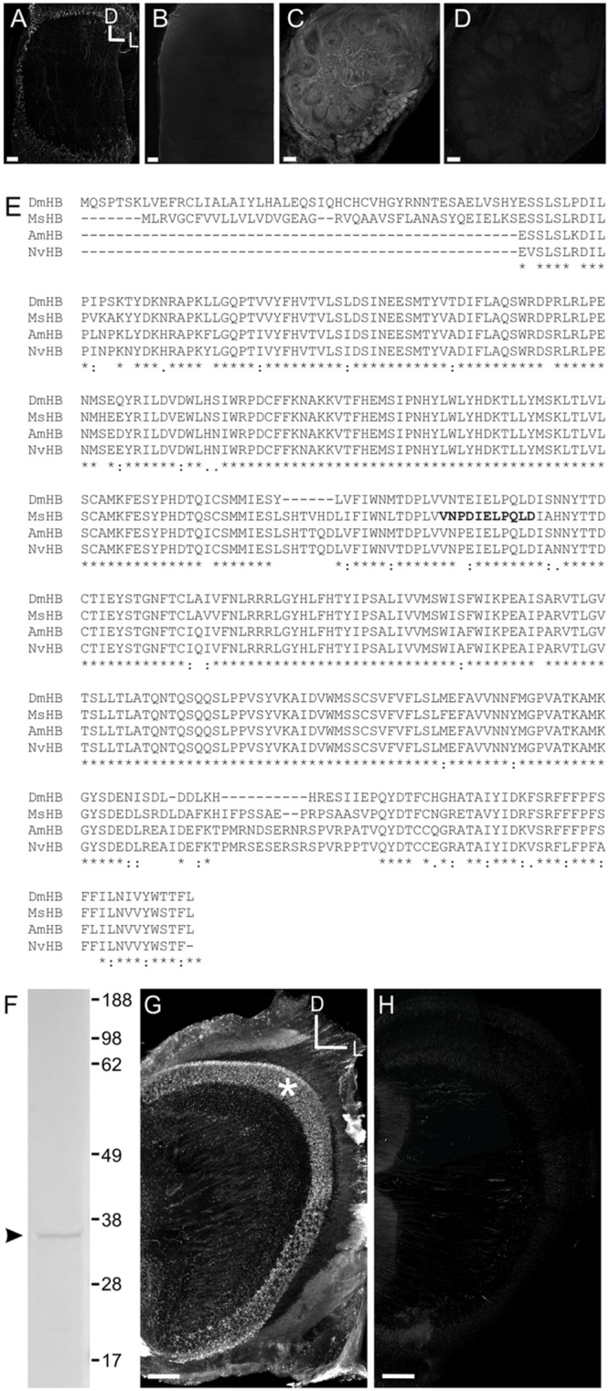
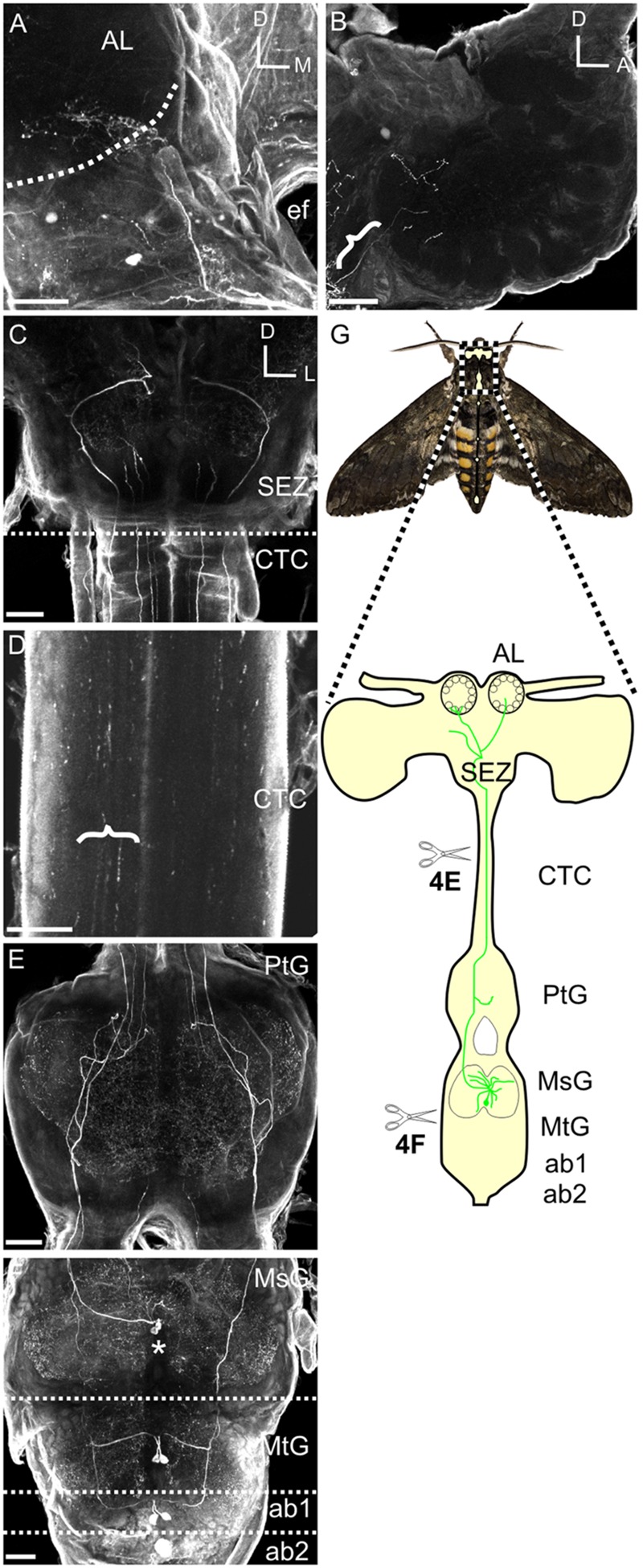
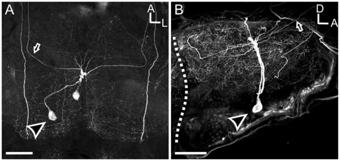
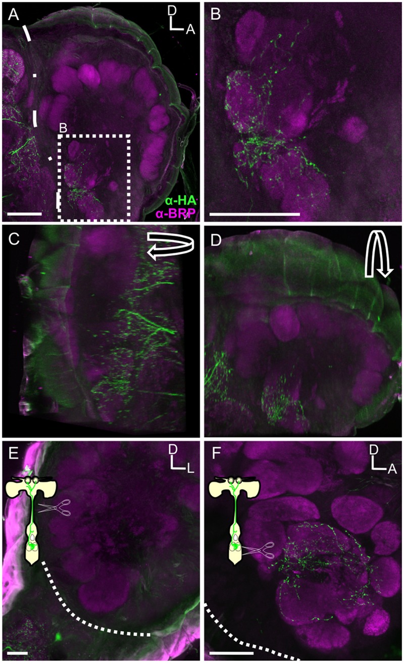
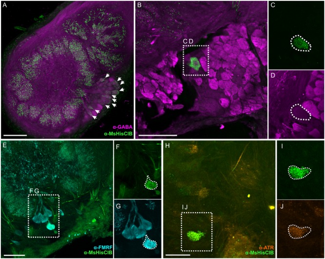
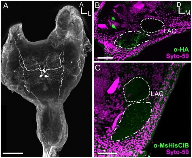
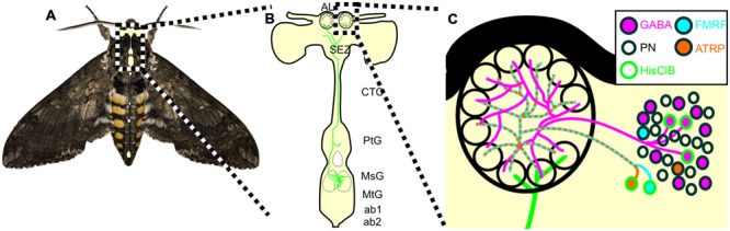
Neural circuits projecting information from motor to sensory pathways are common across sensory domains. These circuits typically modify sensory function as a result of motor pattern activation; this is particularly so in cases where the resultant behavior affects the sensory experience or its processing. However, such circuits have not been observed projecting to an olfactory pathway in any species despite well characterized active sampling behaviors that produce reafferent mechanical stimuli, such as sniffing in mammals and wing beating in the moth Manduca sexta. In this study we characterize a circuit that connects a flight sensory-motor center to an olfactory center in Manduca. This circuit consists of a single pair of histamine immunoreactive (HA-ir) neurons that project from the mesothoracic ganglion to innervate a subset of ventral antennal lobe (AL) glomeruli. Furthermore, within the AL we show that the M. sexta histamine B receptor (MsHisClB) is exclusively expressed by a subset of GABAergic and peptidergic LNs, which broadly project to all olfactory glomeruli. Finally, the HA-ir cell pair is present in fifth stage instar larvae; however, the absence of MsHisClB-ir in the larval antennal center indicates that the circuit is incomplete prior to metamorphosis and importantly prior to the expression of flight behavior. Although the functional consequences of this circuit remain unknown, these results provide the first detailed description of a circuit that interconnects an olfactory system with motor centers driving flight behaviors including odor-guided flight.

Keywords: flight; histamine; insect; modulation; olfaction.


Figures

FIGURE 1
FIGURE 1
Characterization of the HA GABA and Manduca sexta HA B receptor (MsHisClB) antibodies. (A) HA labeling in control animals where the antibody was not pre-adsorbed. HA labeling in the optic lobe which is the primary neurotransmitter used by arthropod receptor cells. (B) HA labeling in the optic lobe is abolished after the HA antibody was pre-adsorbed with a 10:1 HA to antibody solution. (C) GABA labeling remains in control animals where the antibody was not pre-adsorbed with GABA. (D) GABA labeling in the AL is abolished after the GABA antibody was pre-adsorbed with a 10:1 GABA to antibody solution. For each panel the same dorsal lateral axis is used. (E) Amino acid sequence alignment of the HA B receptor subunits of Manduca sexta (MsHB; Msex2.04603-RA), Drosophila melanogaster (DmHB; ACA13298.1), Apis mellifera (AmHB; ABG75740.1), and Nasonia vitripennis (NvHB; ACZ51422.1). Asterisks indicate sequence identity across all four species. Bold font indicates the immunogenic peptide sequence from Manduca that was used to generate the MsHisClB antibody. (F) Western blot using MsHisClB receptor antibody on Manduca brain tissue resulted in a single band at the predicted molecular weight (36 kDa) of the MsHisClB protein. (G) Frontal section of optic lobe depicting MsHisClB-ir in the lamina (as labeled by an asterisks). (H) Pre-adsorption with the immunogenic peptide sequence eliminates all labeling in the lamina. Scale bars = 50 μm. D, dorsal, L, lateral, A, anterior.
FIGURE 2
FIGURE 2
Mesothoracic deutocerebrum histamine neurons project from the MsG to the AL of Manduca sexta. (A) Frontal view of HA-ir labeling in a whole mount brain preparation. Hatched line outlines the AL boundary. (B) Saggital view of a HA-ir process entering the AL (bracket). (C) Frontal view of HA-ir processes entering the SEZ from the cervicothoracic connective. Notice that four pairs enter the SEZ. (D) HA-ir processes in the cervicothoracic connective. Brackets highlight three HA-ir processes. (E) Horizontal view of the HA-ir processes in the prothoracic ganglion. Notice four pairs ascending from here as well. (F) Horizontonal view of HA-ir in the MsG, the metathoracic ganglion, and the first two abdominal ganglia. Each SEZment has a pair of HA-ir cell bodies located in the medial third of their respective ganglion. Asterisks highlights MDNn cell bodies. (G) Schematic of the Manduca nervous system highlighting the MDHns (green). Hatched boundary indicates the MsG. All scale bars = 100 μm. AL, antennal lobe; ef, esophageal foramen; SEZ, subesophageal zone; CTC, cervicothoracic connective; PtG, prothoracic ganglion; MsG, mesothoracic ganglion; MtG, metathoracic ganglion; ab1, abdominal ganglion 1; ab2, abdominal ganglion 2.
FIGURE 3
FIGURE 3
Mesothoracic deutocerebrum histamine neurons processes radiate laterally throughout the MsG, but are primarily restricted to the dorsal aspect. (A) Horizontal view of the MSG showing two cell bodies with each cell projecting out one side of the ganglia. (B) Sagittal section of the MsG shows two large HA-ir cells with cell bodies (black arrow head with a white outline) situated ventrally and a radiating dendritic field dorsally with the axon (black arrow with white outline) projecting up the connective between the mesothoracic and prothoracic ganglia. White dotted line indicates the boundary between the mesothoracic and metathoracic ganglia. Arrow indicates MDHn cell body in each image. All scale bars = 100 μm.
FIGURE 4
FIGURE 4
The MDH neurons provide the sole source of HA-ir input to the ALs. (A) Saggital section of the AL with HA-ir (green). BRP (magenta) outlines glomeruli of the AL. Dotted line outlines the posterior boundary of the AL. Scale bar = 100 μm. (B) High magnification view of inset from (A). Highly varicose HA-ir processes innervate 4–6 ventral posterior glomeruli. Scale bar = 50 μm. (C) Rotation of image (A) about the y-axis showing HA still overlapping with BRP labeling. (D) Rotation of image (A) about the x-axis again showing HA overlapping with BRP labeling, collectively confirming that HA ramifies glomeruli. (E) Frontal section showing that HA-ir is absent in the AL following ablation of the cervicothoracic connective. Scale bar = 100 μm. (F) Sagittal view of HA-ir in the AL following ablation between the MsG and the metathoracic ganglia in which the lesioning of metathoric HA-ir neuron axons was confirmed. Dashed lines indicate boundary of AL in (E,F). Scale bars = 50 μm.
FIGURE 5
FIGURE 5
Within the AL the MsHisClB receptor is expressed by a subset of GABAergic LNs and one FMRF-amidergic and one ATRergic LN. (A) MsHisClB (green) and GABA (magenta) co-labeling in the lateral cell cluster of the AL. MsHisClB-ir is expressed in all AL glomeruli. Scale bar 100 μm. (B) GABA-ir and MsHisClB-ir expression in the lateral cell cluster. (C,D) Inset from (B) highlights a single large MsHisClB-ir cell body that does not express GABA. (E) FMRFamide-ir (cyan) and MsHisClB-ir (green) expression in the lateral cell cluster. (F,G) Inset from (E) highlights a single large cell body that expresses both MsHisClB-ir and FMRFamide-ir. (H) ATR-ir (orange) and MsHisClB-ir (green) expression in the lateral cell cluster. (I,J) Inset from (H) highlights a single large cell body that expresses both MsHisClB-ir and FMRFamide-ir. All scale bars = 50 μm unless otherwise noted.
FIGURE 6
FIGURE 6
The MDHns survive metamorphosis, but the MsHisClB receptor is not expressed in the LAC. (A) Horizontal view of HA-ir in the fifth instar larval MsG shows highly similar cell morphology and radiation patterns of fine processes as in the adult MsG. (B) HA-ir in the larval brain (green) shows extensive branching in the tritocerebrum (dash-dot line), but very little innervation in the LAC (dashed line). Syto-59 (magenta) highlights the boundary of the tritocerebrum and LAC. (C) MsHisClB-ir (green) is present in the tritocerebrum, but not in the LAC. LAC and tritocerebrum highlighted with Syto-59 (magenta) as in (B). All scale bars = 100 μm.
FIGURE 7
FIGURE 7
Schematic of the proposed MDHn circuit. (A) Manduca with overlaid nervous system cartoon. (B) Schematic of the MDHn cells from the thoracic ganglia to the AL. Only one cell is shown in detail with processes radiating in the MsG, a small process in the prothoracic ganglion, projecting up the cervicothoracic connective, a branch to the AMMC, and bilateral projections to each AL. (C) MDHn projection entering the ventral AL (green) along with the proposed AL circuitry. For the sake of simplicity, only the processes from MsHisClB-ir expressing neurons (green outline) are shown. MsHisClB-ir GABAergic (pink with green outline) and peptidergic (cyan or orange with green outline for FMRFamide and ATR, respectively) LNs ramify each glomerulus. Other cell types are also present including PNs (open circles), GABAergic LNs (pink circles with black outlines), ATR LNs (orange circles with black outline), and FMRF LNs (blue circles with black outline). AL, antennal lobe; oe, esophageal foramen; SEZ, subesophageal zone; CTC, cervicothoracic connective; PtG, prothoracic ganglion; MsG, mesothoracic ganglion; MtG, metathoracic ganglion; ab1, abdominal ganglion 1; ab2, abdominal ganglion 2.

References

    1. Adrian E. D. (1942). Olfactory reactions in the brain of the hedgehog. J. Physiol. 100 459–473. 10.1113/jphysiol.1942.sp003955 - DOI - PMC - PubMed
    1. Ando N., Wang H., Shirai K., Kanzaki R. (2011). Central projections of the wing afferents in the hawkmoth, Agrius convolvuli. J. Insect. Physiol. 57 1518–1536. 10.1016/j.jinsphys.2011.08.002 - DOI - PubMed
    1. Baker T. C., Carde R. T. (1979). Analysis of pheromone-mediated behaviors in male Grapholitha molesta, the oriental fruit moth (Lepidoptera: Tortricidae). Environ. Entomol. 8 956–968. 10.1093/ee/8.5.956 - DOI
    1. Bell R. A., Joachim F. G. (1976). Techniques for rearing laboratory colonies of tobacco hornworms and pink bollworms. Ann. Entomol. Soc. Am. 69 365–373. 10.1093/aesa/69.2.365 - DOI
    1. Brainard M. S., Doupe A. J. (2000). Interruption of a basal ganglia-forebrain circuit prevents plasticity of learned vocalizations. Nature 404 762–766. 10.1038/35008083 - DOI - PubMed

Publication types

MeSH terms

LinkOut - more resources