. 2016 Feb 16:7:40.
doi: 10.3389/fimmu.2016.00040. eCollection 2016.

The Non-structural Protein 5 and Matrix Protein Are Antigenic Targets of T Cell Immunity to Genotype 1 Porcine Reproductive and Respiratory Syndrome Viruses

Affiliations

The Non-structural Protein 5 and Matrix Protein Are Antigenic Targets of T Cell Immunity to Genotype 1 Porcine Reproductive and Respiratory Syndrome Viruses

Helen Mokhtar et al. Front Immunol. .

Abstract

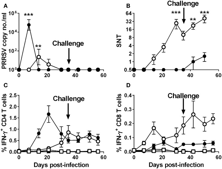
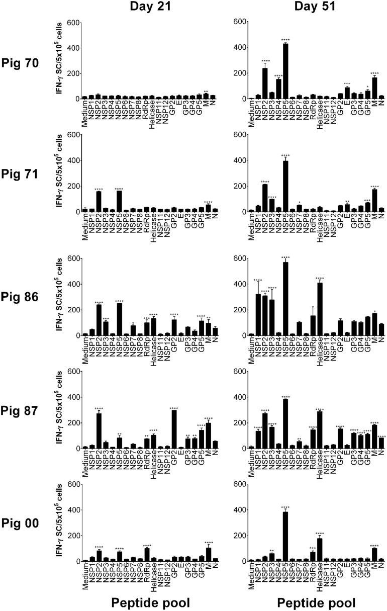
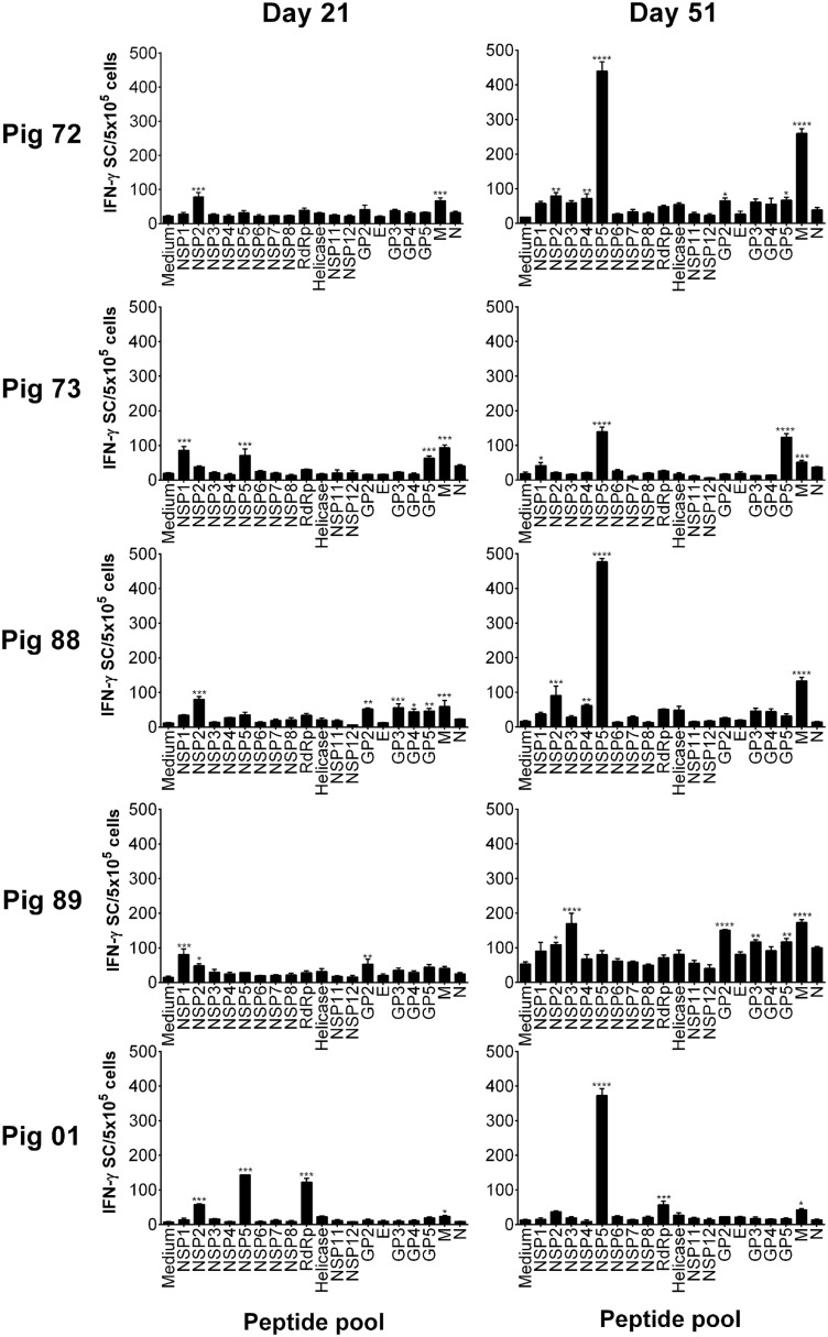
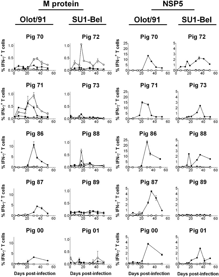
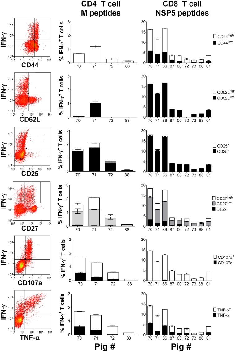
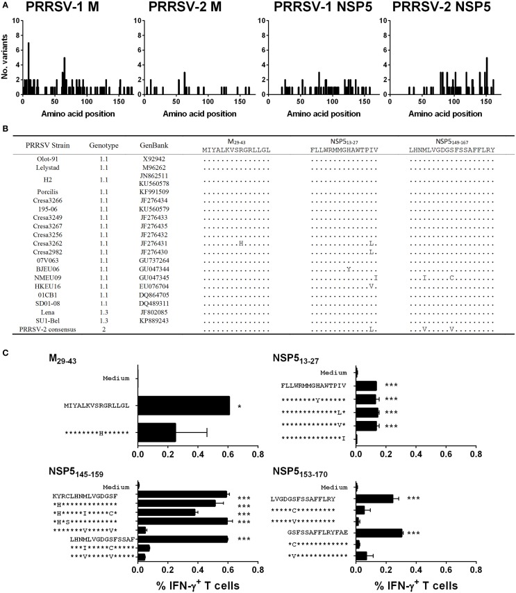
The porcine reproductive and respiratory syndrome virus (PRRSV) is the cause of one of the most economically important diseases affecting swine worldwide. Efforts to develop a next-generation vaccine have largely focused on envelope glycoproteins to target virus-neutralizing antibody responses. However, these approaches have failed to demonstrate the necessary efficacy to progress toward market. T cells are crucial to the control of many viruses through cytolysis and cytokine secretion. Since control of PRRSV infection is not dependent on the development of neutralizing antibodies, it has been proposed that T cell-mediated immunity plays a key role. Therefore, we hypothesized that conserved T cell antigens represent prime candidates for the development a novel PRRS vaccine. Antigens were identified by screening a proteome-wide synthetic peptide library with T cells from cohorts of pigs rendered immune by experimental infections with a closely related (subtype 1) or divergent (subtype 3) PRRSV-1 strain. Dominant T cell IFN-γ responses were directed against the non-structural protein 5 (NSP5), and to a lesser extent, the matrix (M) protein. The majority of NSP5-specific CD8 T cells and M-specific CD4 T cells expressed a putative effector memory phenotype and were polyfunctional as assessed by coexpression of TNF-α and mobilization of the cytotoxic degranulation marker CD107a. Both antigens were generally well conserved among strains of both PRRSV genotypes. Thus, M and NSP5 represent attractive vaccine candidate T cell antigens, which should be evaluated further in the context of PRRSV vaccine development.

Keywords: IFN-γ; T cell; antigen identification; phenotype and function; porcine reproductive and respiratory syndrome virus; vaccine.


Figures

Figure 1
Figure 1
Outcome of infection and subsequent challenge infection with PRRSV-1 strains and association with virus-specific immune responses. Pigs were experimentally infected intranasally with either an attenuated PRRSV-1 strain (Olot/91; open symbols; n = 5) or a virulent sub-genotype 3 strain (SU1-Bel; closed symbols; n = 5) strains on day 0 and day 35 postinfection and PRRSV genome copy numbers in serum assessed (A). Serum neutralizing antibody titers (SNT) were assessed against PRRSV Olot/91 infectivity in vitro (B). IFN-γ expression by CD4+CD8αlow (CD4) and CD4CD8αhigh (CD8) T cells was assessed by flow cytometry following mock (square symbols) or homologous virus (circle symbols) stimulation [(C,D) respectively]. Results are expressed as the mean data for each group, error bars represent the SEM. For RT-qPCR data and VNTs, values were compared to the corresponding value on 0 dpi whereas for T cell IFN-γ data from virus-stimulated cultures were compared against the corresponding mock stimulated culture for each time point using a two-way analysis of variance (ANOVA) followed by a Tukey’s multiple comparison test; ***p < 0.001, **p < 0.01.
Figure 2
Figure 2
Recognition of PRRSV-1 proteins by T cells from pigs experimentally infected with the Olot/91 strain. PBMC from pigs experimentally infected with PRRSV-1 Olot/91 (n = 5) were isolated on day 21 and day 51 postinfection (16 days post-challenge), and stimulated in vitro with synthetic peptides pooled to represent 19 PRRSV-1 proteins. IFN-γ secreting cells were enumerated by ELISpot assay. Data are presented as IFN-γ spot forming cells (SFC)/5 × 105 PBMC (triplicate cultures) for each animal and error bars show the SEM. Values for each peptide pool-stimulated condition were compared to the corresponding unstimulated (medium) control using a one-way ANOVA followed by a Dunnett’s multiple comparison test; ***p < 0.001, **p < 0.01, and *p < 0.05.
Figure 3
Figure 3
Recognition of PRRSV-1 proteins by T cells from pigs experimentally infected with the SU1-Bel strain. PBMC from pigs experimentally infected with PRRSV-1 SU1-Bel (n = 5) were isolated on day 21 and day 51 postinfection (16 days post-challenge), and stimulated in vitro with synthetic peptides pooled to represent 19 PRRSV-1 proteins. IFN-γ secreting cells were enumerated by ELISpot assay. Data are presented as IFN-γ spot forming cells (SFC)/5 × 105 PBMC (triplicate cultures) for each animal and error bars show the SEM. Values for each peptide pool-stimulated condition were compared to the corresponding unstimulated (medium) control using a one-way ANOVA followed by a Dunnett’s multiple comparison test; ***p < 0.001, **p < 0.01, and *p < 0.05.
Figure 4
Figure 4
Assessment of NSP5- and M-specific IFN–γ T cell responses over the course of infection and challenge with PRRSV-1 Olot/91 and SU1-Bel strains. Previously cryopreserved PBMC were stimulated ex vivo with synthetic peptide pools representing M or NSP5 protein or left unstimulated. IFN-γ expression by CD4+CD8αlow (CD4; open symbols) and CD4CD8αhigh (CD8; closed symbols) T cells from unstimulated (square symbols) or peptide pool-stimulated cultures (circle symbols) was assessed by flow cytometry. The mean % of IFN-γ+ T cells from duplicate cultures are presented for each animal and error bars show the SEM. Data from peptide pool-stimulated cultures were compared against the corresponding unstimulated culture using a two-way ANOVA followed by a Tukey’s multiple comparison test.
Figure 5
Figure 5
Assessment of the phenotype and polyfunctionality of PRRSV-1 NSP5-specific CD8 T cells. Previously cryopreserved PBMC from identified T cell responder pigs on day 30 postinfection were stimulated with synthetic peptide pools representing M or NSP5 proteins or left unstimulated. The expression of CD44, CD62L, CD25, CD27, surface CD107a, and TNF-α by IFN-γ+ CD4 T cells in response to M peptides and CD8 T cells in response to NSP5 peptides were assessed by flow cytometry as shown by representative dot plots. The mean % of unstimulated-corrected data from duplicate cultures are presented for individual animals and error bars show the SEM.
Figure 6
Figure 6
Assessment of the conservation of identified T cell antigens M and NSP5 among PRRSV strains and assessment of T cell reactivity against variant peptides. The complete predicted amino acid sequences of M (from 64 PRRSV-1 and 31 PRRSV-2 strains) and NSP5 (from 19 PRRSV-1 and 36 PRRSV-2 strains) were aligned and the number of different amino acid variants at each residue plotted (A). The predicted amino acid sequences of identified antigenic regions, M29-43, NSP513-27, and NSP5149-167, were compared among the panel of 19 PRRSV-1 strains, for which both M and NSP5 sequence data were available, and a consensus sequence based on available PRRSV-2 strains (B). Based on the observed amino acid substitutions, variant peptides were used to stimulate PBMC from pigs 71 and 86 (C). IFN-γ expression by CD4 T cells to M29-43 (pig 71) and CD8 T cells to NSP513-27 (pig 71), NSP5145-159 (pig 86), and NSP5153-170 (pig 71) peptides were assessed by flow cytometry. The mean% of unstimulated (medium) and peptide stimulated data from duplicate cultures are presented and error bars show the SEM. Values were compared to the unstimulated control using a one-way ANOVA followed by a Dunnett’s multiple comparison test; ***p < 0.001, **p < 0.01, and *p < 0.05.

Similar articles

Cited by

References

    1. Holtkamp DJ, Kliebenstein JB, Neumann EJ, Mowrer C, Haley C. Assessment of the economic impact of porcine reproductive and respiratory syndrome virus on United States pork producers. J Swine Health Prod (2013) 21:72–84.
    1. Tong GZ, Zhou YJ, Hao XF, Tian ZJ, An TQ, Qiu HJ. Highly pathogenic porcine reproductive and respiratory syndrome, China. Emerg Infect Dis (2007) 13:1434–6.10.3201/eid1309.070399 - DOI - PMC - PubMed
    1. Feng Y, Zhao T, Nguyen T, Inui K, Ma Y, Nguyen TH, et al. Porcine respiratory and reproductive syndrome virus variants, Vietnam and China, 2007. Emerg Infect Dis (2008) 14:1774–6.10.3201/eid1411.071676 - DOI - PMC - PubMed
    1. Karniychuk UU, Geldhof M, Vanhee M, Van Doorsselaere J, Saveleva TA, Nauwynck HJ. Pathogenesis and antigenic characterization of a new East European subtype 3 porcine reproductive and respiratory syndrome virus isolate. BMC Vet Res (2010) 6:30.10.1186/1746-6148-6-30 - DOI - PMC - PubMed
    1. Morgan SB, Graham SP, Salguero FJ, Sanchez Cordon PJ, Mokhtar H, Rebel JM, et al. Increased pathogenicity of European porcine reproductive and respiratory syndrome virus is associated with enhanced adaptive responses and viral clearance. Vet Microbiol (2013) 163:13–22.10.1016/j.vetmic.2012.11.024 - DOI - PubMed