

Myeloma bone disease: Pathophysiology and management
- PMID: 26909272
- PMCID: PMC4723362
- DOI: 10.1016/j.jbo.2013.04.001
Myeloma bone disease: Pathophysiology and management
Abstract
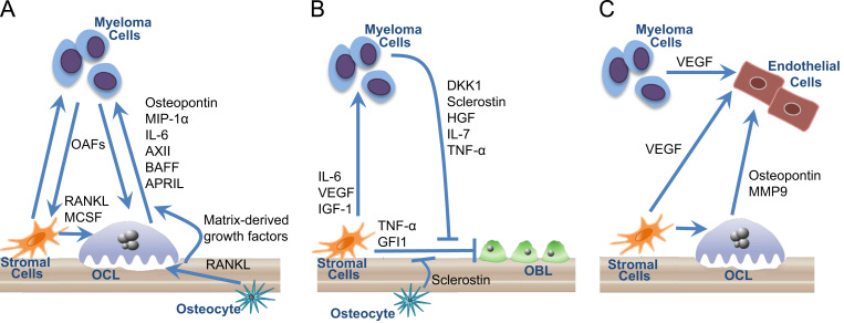
Multiple myeloma bone disease is marked by severe dysfunction of both bone formation and resorption and serves as a model for understanding the regulation of osteoblasts (OBL) and osteoclasts (OCL) in cancer. Myeloma bone lesions are purely osteolytic and are associated with severe and debilitating bone pain, pathologic fractures, hypercalcemia, and spinal cord compression, as well as increased mortality. Interactions within the bone marrow microenvironment in myeloma are responsible for the abnormal bone remodeling in myeloma bone disease. Myeloma cells drive bone destruction that increases tumor growth, directly stimulates the OCL formation, and induces cells in the marrow microenvironment to produce factors that drive OCL formation and suppress OBL formation. Factors produced by marrow stromal cells and OCL promote tumor growth through direct action on myeloma cells and by increasing angiogenesis. Current therapies targeting MMBD focus on preventing osteoclastic bone destruction; however regulators of OBL inhibition in MMBD have also been identified, and targeted agents with a potential anabolic effect in MMBD are under investigation. This review will discuss the mechanisms responsible for MMBD and therapeutic approaches currently in use and in development for the management of MMBD.
Keywords: Angiogenesis; Bone marrow microenvironment; Myeloma; Myeloma bone disease; Osteoblast; Osteoclast.
Figures
References
-
- Roodman GD. Mechanisms of bone metastasis. The New England journal of medicine. 2004;350:1655–1664. - PubMed
-
- Terpos E, Berenson J, Cook RJ, Lipton A, Coleman RE. Prognostic variables for survival and skeletal complications in patients with multiple myeloma osteolytic bone disease. Leukemia: Official Journal of the Leukemia Society of America, Leukemia Research Fund, UK. 2010;24:1043–1049. - PubMed
-
- Melton LJ, 3rd, Kyle RA, Achenbach SJ, Oberg AL, Rajkumar SV. Fracture risk with multiple myeloma: a population-based study. Journal of Bone and Mineral Research: the Official Journal of the American Society for Bone and Mineral Research. 2005;20:487–493. - PubMed
-
- Saad F, Lipton A, Cook R, Chen YM, Smith M, Coleman R. Pathologic fractures correlate with reduced survival in patients with malignant bone disease. Cancer. 2007;110:1860–1867. - PubMed
-
- Diamond T, Levy S, Day P, Barbagallo S, Manoharan A, Kwan YK. Biochemical, histomorphometric and densitometric changes in patients with multiple myeloma: effects of glucocorticoid therapy and disease activity. British Journal of Haematology. 1997;97:641–648. - PubMed
Publication types
LinkOut - more resources
Full Text Sources
Other Literature Sources
Miscellaneous
