. 2015 Oct 20;4(12):916-25.
doi: 10.1016/j.molmet.2015.09.008. eCollection 2015 Dec.

A human beta cell line with drug inducible excision of immortalizing transgenes

Affiliations

A human beta cell line with drug inducible excision of immortalizing transgenes

Marion Benazra et al. Mol Metab. .

Abstract

Objectives: Access to immortalized human pancreatic beta cell lines that are phenotypically close to genuine adult beta cells, represent a major tool to better understand human beta cell physiology and develop new therapeutics for Diabetes. Here we derived a new conditionally immortalized human beta cell line, EndoC-βH3 in which immortalizing transgene can be efficiently removed by simple addition of tamoxifen.

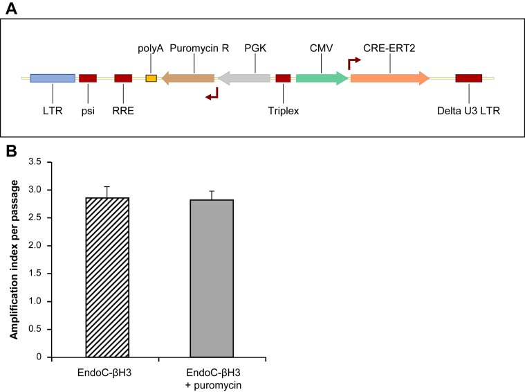
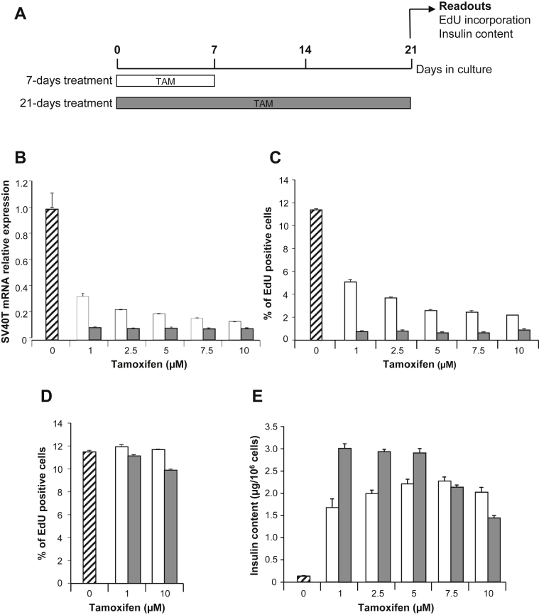
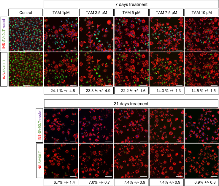
Methods: We used lentiviral mediated gene transfer to stably integrate a tamoxifen inducible form of CRE (CRE-ERT2) into the recently developed conditionally immortalized EndoC βH2 line. The resulting EndoC-βH3 line was characterized before and after tamoxifen treatment for cell proliferation, insulin content and insulin secretion.

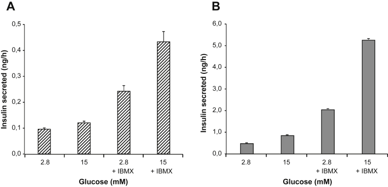
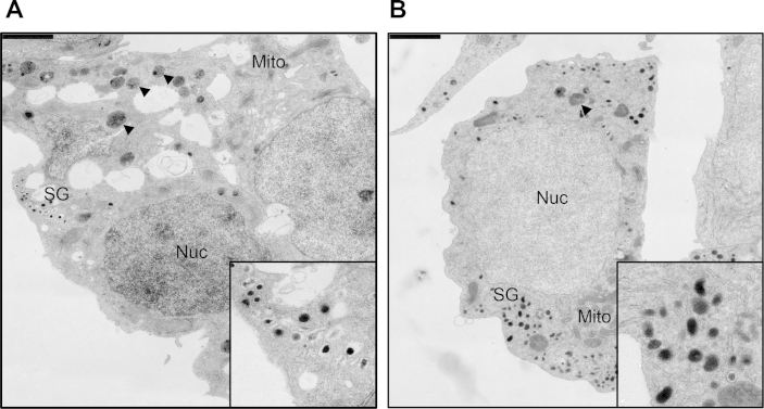
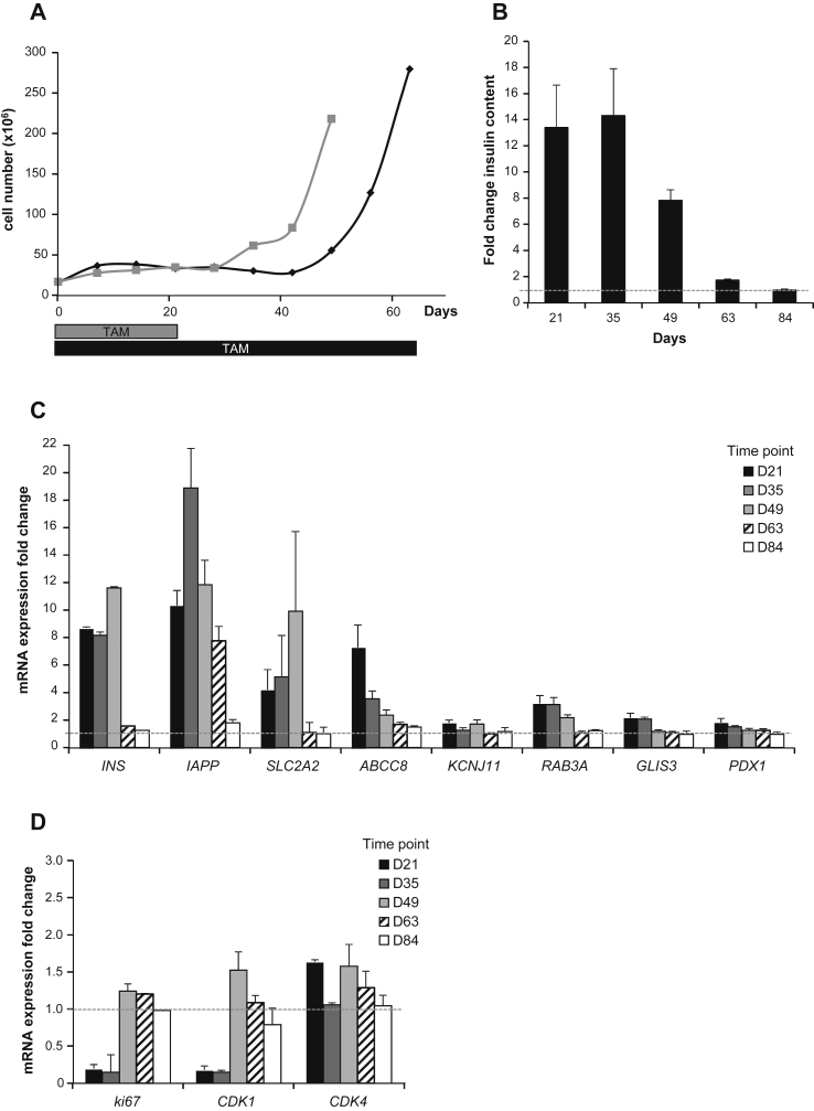
Results: We showed that EndoC-βH3 expressing CRE-ERT2 can be massively amplified in culture. We established an optimized tamoxifen treatment to efficiently excise the immortalizing transgenes resulting in proliferation arrest. In addition, insulin expression raised by 12 fold and insulin content increased by 23 fold reaching 2 μg of insulin per million cells. Such massive increase was accompanied by enhanced insulin secretion upon glucose stimulation. We further observed that tamoxifen treated cells maintained a stable function for 5 weeks in culture.

Conclusions: EndoC βH3 cell line represents a powerful tool that allows, using a simple and efficient procedure, the massive production of functional non-proliferative human beta cells. Such cells are close to genuine human beta cells and maintain a stable phenotype for 5 weeks in culture.

Keywords: Cell engineering; Conditional immortalization; Human beta cell function; Human pancreatic beta cell line; Tamoxifen inducible CRE.


Figures

Figure 1
Figure 1
Production of EndoC-βH3 cells a Tamoxifen inducible excisable human beta cell line derived from EndoC-βH2. (A) Schematic representation of lentiviral vector used to produce EndoC-βH3 cells expressing both the TAM inducible form of CRE (CRE-ERT2) and the resistance to puromycin (B) Cells were passaged every week. Amplification index was defined as fold change between the initial number of seeded cells and the one counted one week later before cell passage. Data represent average of amplification index ± S.E.M. over 10 passages of EndoC-βH3 cells cultured with or without antibiotic (puromycin) selection.
Figure 2
Figure 2
Definition of optimal procedure for efficient Tamoxifen treatment of EndoC-βH3. (A) Schematic representation of the experimental optimization procedure for TAM treatment. (B, C, E) EndoC-βH3 cells were treated for 7 days (white box) or 21 days (Gray box) with TAM. Un-treated cells are represented as hatched box. Proliferation assays and insulin content measurements were performed at day 21. (B) Effect of TAM treatment on SV40LT gene expression. Quantitative PCR results are representative of 2 independent experiments performed in triplicates. Data corresponded to average relative expression values normalized to TPB expression ± S.E.M. (CD) Effect of TAM treatment on EdU incorporation by EndoC-βH3 cells (C) or EndoC-βH2 (D) relative to total cell number. EdU was added for 1 h and analysis was performed by FACS. Results are representative of 2 independent experiments performed in duplicates. Data are expressed as average percentage ± S.E.M. (E) Effect of TAM treatment on insulin content in EndoC-βH3. Data are expressed as insulin content per million cells ± S.E.M. of 3 independent wells per condition assayed in triplicates by ELISA.
Figure 3
Figure 3
SV40LT and insulin immunostaining following Tamoxifen treatment. SV40LT (green), Insulin (red) Nuclei (blue) immunofluorescent staining of control or excised cells analyzed 21 days after a 7 or 21 days TAM treatment. Percent of SV40LT positive nuclei relative to total number of nuclei is indicated below each conditions. Confocal acquisition settings were identical for all conditions. Scale bar = 50 μm.
Figure 4
Figure 4
Glucose responsive insulin secretion in EndoC-βH3 following treatment with Tamoxifen. EndoC-βH3 were treated or not for 21 days with 1 μM TAM. (A) Glucose stimulated insulin secretion (GSIS) on un-treated EndoC-βH3 in presence or absence of IBMX. (B) GSIS on TAM-treated EndoC-βH3 in presence or absence of IBMX. (AB) GSIS data are expressed as ng of secreted insulin per hour ± S.E.M. of 3 independent wells seeded with 7 × 104 cells per condition assayed in triplicates by ELISA.
Figure 5
Figure 5
TEM analysis of treated and untreated EndoC-βH3. Non-treated and treated EndoC-βH3 cells were analyzed after Epon embedding and ultrathin sectioning. (A) Non-treated EndoC-βH3, inset: detail of insulin SGs, (B) TAM-treated EndoC-βH3, inset: detail of insulin SGs. Scale bar: 2 μm. Labeling key: Nuc, nucleus; Mito, mitochondria; SG, secretory granules; arrowheads, degradative compartments.
Figure 6
Figure 6
Long-term phenotype of Tamoxifen treated EndoC-βH3. (A) Cell growth monitoring over time (every 7 days) during 63 days. TAM treatment was either stopped at day 21 (in gray), or continued during the whole experiment (in black). (B) Insulin content was determined in EndoC-βH3 cells continuously treated with TAM and compared to untreated cells. Analysis was performed every second week, starting 21 days after initial TAM treatment. Data are expressed as fold change over untreated cells (dotted line) of insulin content ± S.E.M. of 3 independent wells per condition assayed in replicates by ELISA. (CD) Gene expression by quantitative RT-PCR was determined in EndoC-βH3 cells that were continuously treated with TAM and compared to untreated cells. Analysis was performed every second week, starting 21 days after initial TAM treatment. Data are expressed as fold change of mRNA expression relative to TBP ± S.E.M. of 3 independent RNA preparations assayed in triplicates.

References

    1. Scharfmann R., Rachdi L., Ravassard P. Concise review: in search of unlimited sources of functional human pancreatic beta cells. Stem Cells Translational Medicine. 2013;2(1):61–67. - PMC - PubMed
    1. Nair G., Hebrok M. Islet formation in mice and men: lessons for the generation of functional insulin-producing beta-cells from human pluripotent stem cells. Current Opinion In Genetics & Development. 2015;32:171–180. - PMC - PubMed
    1. Newby B.N., Terada N., Mathews C.E. In search of a surrogate: engineering human beta cell lines for therapy. Trends in Endocrinology and Metabolism. 2014;25(8):378–380. - PMC - PubMed
    1. Kroon E., Martinson L.A., Kadoya K., Bang A.G., Kelly O.G., Eliazer S. Pancreatic endoderm derived from human embryonic stem cells generates glucose-responsive insulin-secreting cells in vivo. Nature Biotechnology. 2008;26(4):443–452. - PubMed
    1. D'Amour K.A., Bang A.G., Eliazer S., Kelly O.G., Agulnick A.D., Smart N.G. Production of pancreatic hormone-expressing endocrine cells from human embryonic stem cells. Nature Biotechnology. 2006;24(11):1392–1401. - PubMed