. 2015;52(5):306-20.
doi: 10.1159/000443884. Epub 2016 Feb 25.

Calreticulin Regulates Neointima Formation and Collagen Deposition following Carotid Artery Ligation

Affiliations

Calreticulin Regulates Neointima Formation and Collagen Deposition following Carotid Artery Ligation

Kurt A Zimmerman et al. J Vasc Res. 2015.

Abstract

Background/aims: The endoplasmic reticulum (ER) stress protein, calreticulin (CRT), is required for the production of TGF-β-stimulated extracellular matrix (ECM) by fibroblasts. Since TGF-β regulates vascular fibroproliferative responses and collagen deposition, we investigated the effects of CRT knockdown on vascular smooth-muscle cell (VSMC) fibroproliferative responses and collagen deposition.

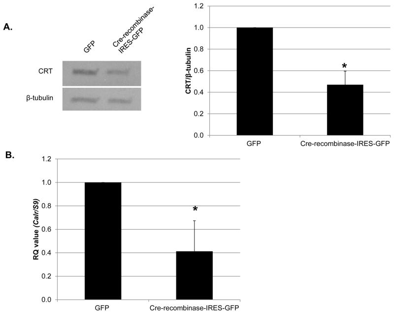
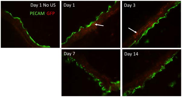
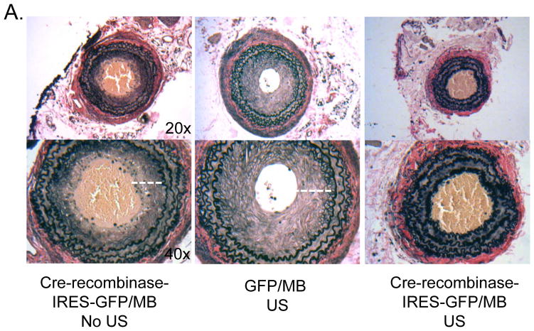
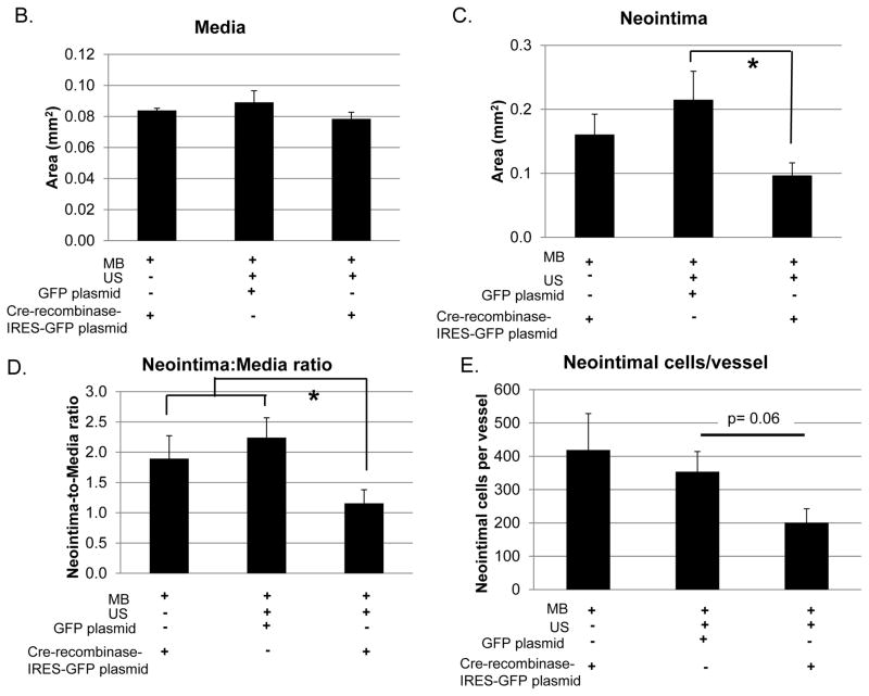
Methods: Using a carotid artery ligation model of vascular injury, Cre-recombinase-IRES-GFP plasmid was delivered with microbubbles (MB) to CRT-floxed mice using ultrasound (US) to specifically reduce CRT expression in the carotid artery.

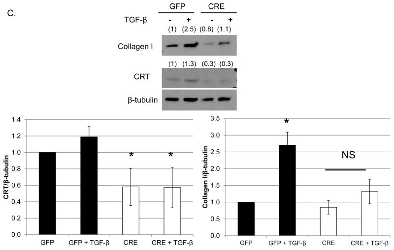
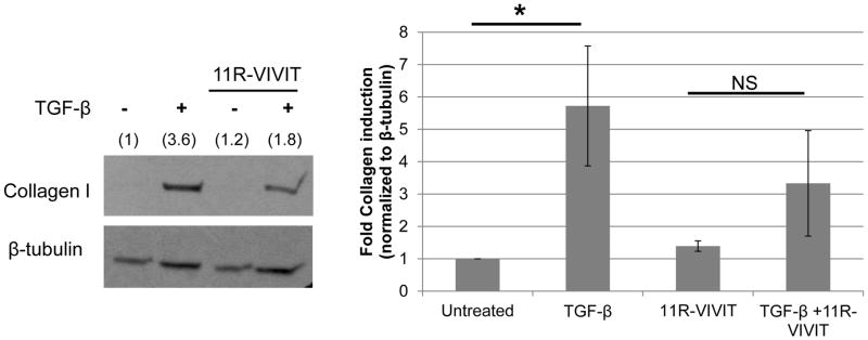
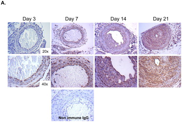
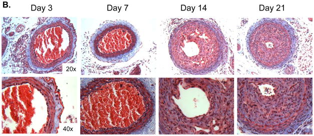
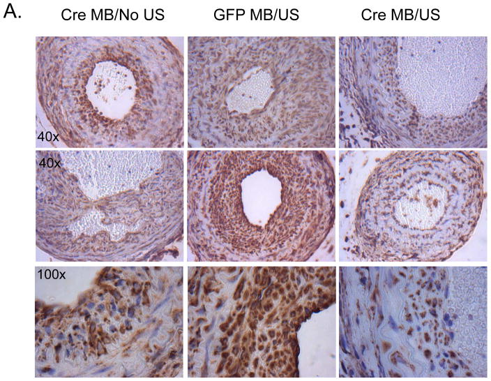
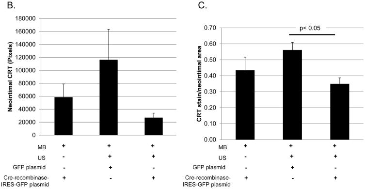
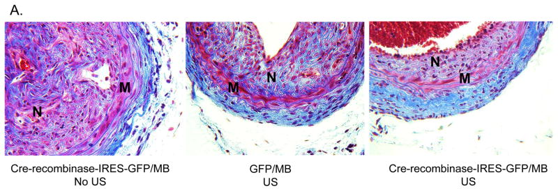
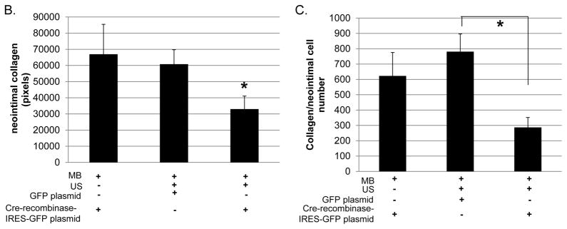
Results: In vitro, Cre-recombinase-mediated CRT knockdown in isolated, floxed VSMCs decreased the CRT transcript and protein, and attenuated the induction of collagen I protein in response to TGF-β. TGF-β stimulation of collagen I was partly blocked by the NFAT inhibitor 11R-VIVIT. Following carotid artery ligation, CRT staining was upregulated with enhanced expression in the neointima 14-21 days after injury. Furthermore, Cre-recombinase-IRES-GFP plasmid delivered by targeted US reduced CRT expression in the neointima of CRT-floxed mice and led to a significant reduction in neointima formation and collagen deposition. The neointimal cell number was also reduced in mice, with a local, tissue-specific knockdown of CRT.

Conclusions: This work establishes a novel role for CRT in mediating VSMC responses to injury through the regulation of collagen deposition and neointima formation.


Conflict of interest statement

Disclosures: There are no conflicts of interest.

Figures

Figure 1
Figure 1. Knockdown of CRT in VSMCs isolated from CRT floxed mice inhibits TGF-β stimulated collagen production
(A, B) VSMCs were isolated from CRT floxed mice, transfected with 1 μg GFP or cre-recombinase-IRES-GFP, and grown overnight in media with 10% FBS. Cells were switched to serum free DMEM for the remainder of the experiment. (A) After 96 total hours, cell lysates were immunoblotted for CRT and normalized to β-tubulin. (n=3 separate experiments). A representative blot is shown in (A). (B) After 72 total hours, RNA was harvested by TRIZOL and transcript levels of Calr and S9 were determined by quantitative real time PCR (n=3 separate experiments). (C) CRT floxed VSMCs were transfected with 1 μg GFP or cre-recombinase-IRES-GFP plasmid, grown overnight in DMEM with 10% FBS, and then switched to serum free DMEM for 24 hours. Cells were treated with 100 pM TGF-β for 48 hours and cell lysates immunoblotted for CRT and type I collagen. A representative blot is shown. Densitometric analyses represent mean density normalized to β-tubulin (indicated above the bands) +/− S.D. (n=3 separate experiments). *p<0.05 vs GFP transfected cells.
Figure 2
Figure 2. CRT regulates TGF-β stimulated collagen production in VSMCs through an NFAT-dependent manner
(A) CRT floxed VSMCs were grown overnight in DMEM with 10% FBS, switched to serum free DMEM overnight, and pretreated with 1 μM 11R-VIVIT for 60 minutes prior to adding 100 pM TGF-β. Cells were then treated daily with TGF-β +/− 11R-VIVIT peptide over 48 hours. Cell lysates were immunoblotted for type I collagen. A representative blot is shown. Bands were analyzed by densitometry and normalized to β-tubulin (indicated above band). Results are mean density normalized to β-tubulin +/− S.D. from 3 separate experiments. *p<0.05 vs untreated cells.
Figure 3
Figure 3. CRT staining and fibrillary collagens are increased in the neointima following carotid artery ligation
(A, B) Carotid artery ligation was performed on CRT floxed mice and carotid arteries were harvested 3, 7, 14, and 21 days following ligation. (A) Immunostaining for CRT was performed using a rabbit monoclonal anti-CRT antibody. Representative images depict strong CRT staining in the neointima 14 and 21 days post ligation. The original magnification of the images is 20x (top panels) and 40x (bottom panels). A control panel is shown from an artery harvested at 7 days in which non-immune rabbit IgG was used as the primary antibody. (B) Representative Masson’s Trichrome stained cross sections of right carotid arteries harvested 3, 7, 14, and 21 days following ligation are shown. The original magnification of the images is 20x (top panels) and 40x (bottom panels).
Figure 4
Figure 4. Delivery of GFP plasmid to medial cells by targeted ultrasound
CRT floxed mice were injected via tail vein with 300 μg GFP plasmid with MB, subjected to US and carotid arteries harvested 1, 3, 7, and 14 days following US. Injection of GFP plasmid with MB but lacking US and harvested at day 1 served as a control. Immunofluorescence for GFP (red) and PECAM1 (green) was performed on longitudinal sections and images taken at 40x. White arrows indicate individual smooth muscle cells which were transfected with the GFP plasmid. Representative images are shown.
Figure 5
Figure 5. Delivery of Cre-recombinase plasmid to the carotid artery of CRT floxed mice reduces neointimal CRT levels
CRT floxed mice with treated with carotid artery-targeted US in the presence of MB to deliver Cre-recombinase-IRES-GFP or GFP plasmids. Cre-recombinase plasmid delivered with MB but without US (Cre MB/No US) served as an additional control. Twenty one days following carotid artery ligation, carotid arteries were perfusion fixed, embedded in paraffin, and sectioned. (A) Immunostaining for CRT was performed using the rabbit monoclonal anti-CRT antibody. Initial magnification was 40x (top, middle panel) or 100x (bottom panel). Control sections in which non-immune rabbit IgG was substituted for the primary antibody were negative (data not shown). (B, C) CRT stain was quantified using Metamorph Imaging Software: (B) total CRT stain (pixels) in the neointima (C) CRT stain normalized to neointima area. Results are expressed as mean values +/− SEM (n=3, cre-recombinase-IRES-GFP with MB/NO US; n=7, GFP with MB/US; or n=7, Cre-recombinase-IRES-GFP with MB/US.) *p<0.05 Cre-recombinase with MB/US vs GFP plasmid with MB/US.
Figure 6
Figure 6. Neointima formation following carotid ligation is reduced in mice treated with Cre-recombinase with MB and US
Carotid arteries were harvested 21 days following ligation and subjected to analyses. (A) Representative elastin stained cross sections of right carotid arteries (original magnification 20x top panel, 40x bottom panel). Quantification of medial area (B), neointimal area (C), and neointima-to-media ratio (D) are shown. Neointima is indicated by a white dashed line in the control animals (40x images). The Cre-recombinase MB/US image (right) does not have a measurable neointima. Clots are observed in the lumens of the Cre-recombinase MB and MB/US arteries. Images quantified using NIH Image J software were obtained using a 20X objective. (E) Quantification of neointimal cell number per vessel 3 weeks following carotid artery ligation is shown. Total nuclei were counted using the 500 μm H&E stained section. Results are expressed as means +/− SEM (n=3,cre-recombinase-IRES-GFP with MB/NO US; n=7, GFP with MB and US; or n=7, Cre-recombinase-IRES-GFP with MB and US.) *p<0.05.
Figure 7
Figure 7. Neointimal collagen is reduced in mice treated with Cre-recombinase plasmid with MB and US
(A) Representative Masson’s Trichrome stained cross sections of right carotid arteries 3 weeks following carotid artery ligation. (B) Quantification of total neointimal collagen content and (C) collagen content normalized to neointimal cell number were performed using Metamorph Imaging Software. Images were obtained using a 40x objective. “M” represents media and “N” represents neointima. Results are expressed as means +/− SEM (n=3, cre-recombinase-IRES-GFP with MB/NO US; n=7, GFP with MB and US; or n=7, Cre-recombinase-IRES-GFP with MB and US.) *p<0.05

Similar articles

Cited by

References

    1. Hoffmann R, Mintz GS, Dussaillant GR, Popma JJ, Pichard AD, Satler LF, Kent KM, Griffin J, Leon MB. Patterns and mechanisms of in-stent restenosis. A serial intravascular ultrasound study. Circulation. 1996;94:1247–1254. - PubMed
    1. Majesky MW. Neointima formation after acute vascular injury. Role of counteradhesive extracellular matrix proteins. Texas Heart Institute journal / from the Texas Heart Institute of St Luke’s Episcopal Hospital, Texas Children’s Hospital. 1994;21:78–85. - PMC - PubMed
    1. Chaabane C, Otsuka F, Virmani R, Bochaton-Piallat ML. Biological responses in stented arteries. Cardiovasc Res. 2013;99:353–363. - PubMed
    1. Majesky MW, Lindner V, Twardzik DR, Schwartz SM, Reidy MA. Production of transforming growth factor beta 1 during repair of arterial injury. J Clin Invest. 1991;88:904–910. - PMC - PubMed
    1. Nabel EG, Shum L, Pompili VJ, Yang ZY, San H, Shu HB, Liptay S, Gold L, Gordon D, Derynck R, et al. Direct transfer of transforming growth factor beta 1 gene into arteries stimulates fibrocellular hyperplasia. Proc Natl Acad Sci U S A. 1993;90:10759–10763. - PMC - PubMed

Publication types

MeSH terms