. 2016 Jun;15(3):499-508.
doi: 10.1111/acel.12456. Epub 2016 Feb 22.

Acetylation reduces SOX9 nuclear entry and ACAN gene transactivation in human chondrocytes

Affiliations

Acetylation reduces SOX9 nuclear entry and ACAN gene transactivation in human chondrocytes

Michal Bar Oz et al. Aging Cell. 2016 Jun.

Erratum in

  • Corrigendum.
    [No authors listed] [No authors listed] Aging Cell. 2017 Aug;16(4):900. doi: 10.1111/acel.12609. Aging Cell. 2017. PMID: 28699313 Free PMC article. No abstract available.

Abstract

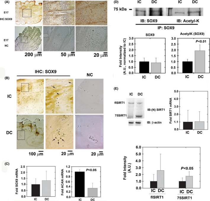
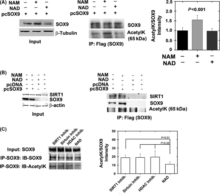
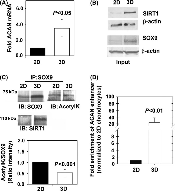
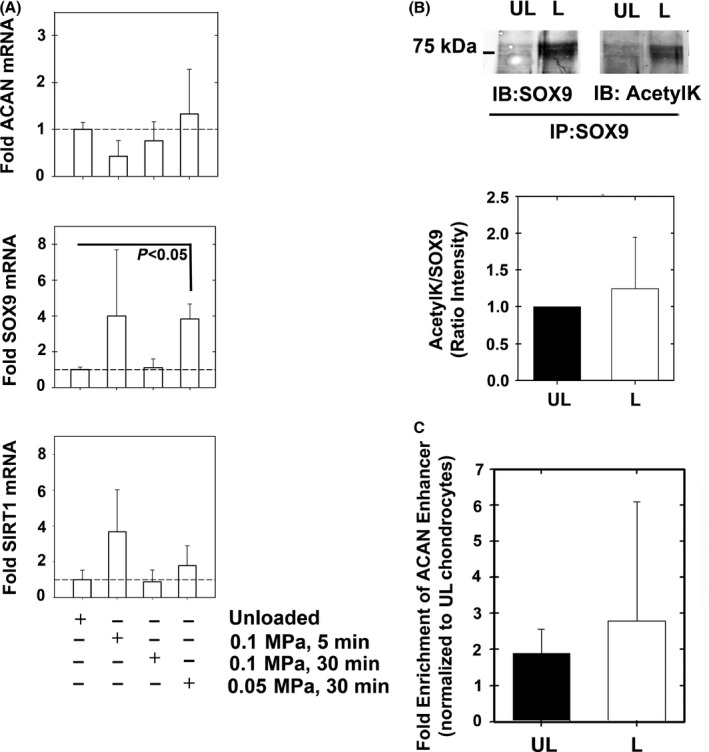
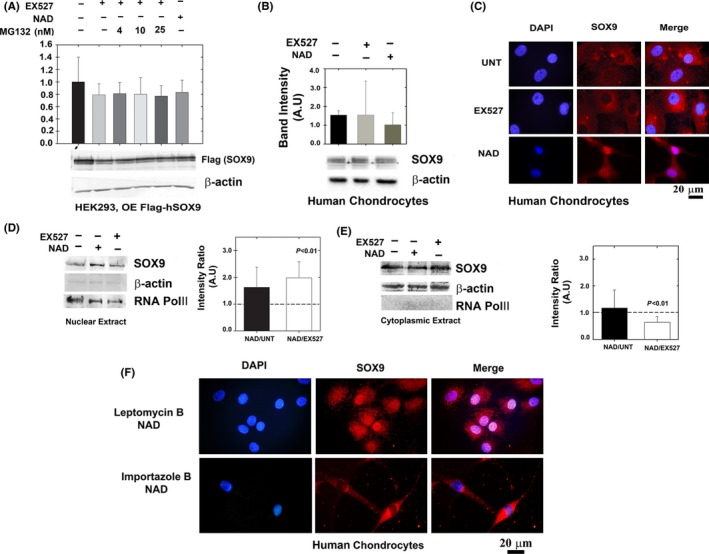
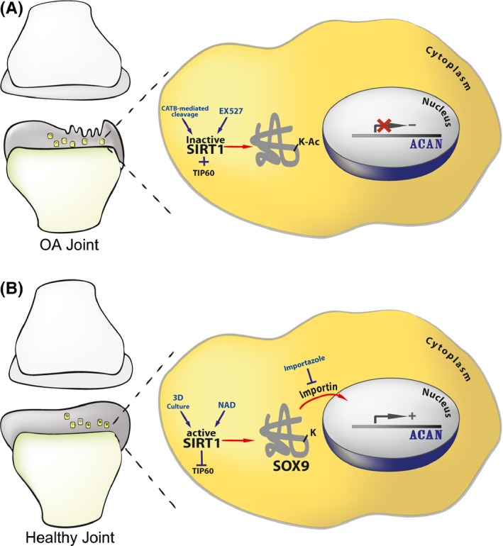
Changes in the content of aggrecan, an essential proteoglycan of articular cartilage, have been implicated in the pathophysiology of osteoarthritis (OA), a prevalent age-related, degenerative joint disease. Here, we examined the effect of SOX9 acetylation on ACAN transactivation in the context of osteoarthritis. Primary chondrocytes freshly isolated from degenerated OA cartilage displayed lower levels of ACAN mRNA and higher levels of acetylated SOX9 compared with cells from intact regions of OA cartilage. Degenerated OA cartilage presented chondrocyte clusters bearing diffused immunostaining for SOX9 compared with intact cartilage regions. Primary human chondrocytes freshly isolated from OA knee joints were cultured in monolayer or in three-dimensional alginate microbeads (3D). SOX9 was hypo-acetylated in 3D cultures and displayed enhanced binding to a -10 kb ACAN enhancer, a result consistent with higher ACAN mRNA levels than in monolayer cultures. It also co-immunoprecipitated with SIRT1, a major deacetylase responsible for SOX9 deacetylation. Finally, immunofluorescence assays revealed increased nuclear localization of SOX9 in primary chondrocytes treated with the NAD SIRT1 cofactor, than in cells treated with a SIRT1 inhibitor. Inhibition of importin β by importazole maintained SOX9 in the cytoplasm, even in the presence of NAD. Based on these data, we conclude that deacetylation promotes SOX9 nuclear translocation and hence its ability to activate ACAN.

Keywords: Aging; SIRT1; SOX9; acetylation; aggrecan; cartilage; nucleus; osteoarthritis.


Figures

Figure 1
Figure 1
SOX9 levels in healthy and diseased articular cartilage. (A) Sagittal sections of E17 hind paws served as positive and negative controls (NC) for immunohistochemistry (IHC) protocols to detect SOX9 protein levels (n = 5).(B) OA‐derived intact cartilage (IC) and degenerated cartilage (DC) were analyzed for SOX9 levels using immunohistochemistry (n = 10). (C) SOX9 and ACAN mRNA levels of freshly isolated chondrocytes from IC and DC samples (n = 8). As indicated in the Materials and methods section, GAPDH was used as housekeeping gene. (D) SOX9 from IC and DC samples was immunoprecipitated and blotted for SOX9 and acetyl‐lysine (AcetylK; n = 5). (E) Freshly isolated chondrocytes from IC and DC cartilage were analyzed with an N‐terminally reactive antibody for SIRT1 to identify its cleavage (n = 9). Full‐length SIRT1 is denoted as flSIRT1, while 75SIRT1 is the 75 kDa cleaved variant generated by cathepsin B‐mediated cleavage of SIRT1 on its C‐terminal domain. Right graphs shows mRNA expression for SIRT1 in freshly isolated chondrocytes from IC and DC samples (n = 8). Image J was employed to quantify band intensity of all immunoblots. SOX9 acetylation was calculated based on the intensity of acetyl‐lysine (AcetylK) band divided by the band intensity of normalized SOX9, assuming the immunoprecipitate consists mainly of SOX9. Statistical significance was determined based on Mann–Whitney U‐test, assuming a P < 0.05 to be statistically significant.
Figure 2
Figure 2
SIRT1 binds and predominantly deacetylates SOX9. HEK293 cells were transfected with a SOX9 expression plasmid (pcSOX9) or pcDNA control and incubated for 24 h with NAD (cofactor of sirtuins) or NAM (a noncompetitive inhibitor of sirtuins). (A) Following immunoblotting for inputs to verify SOX9 expression (right panel), the extracts were immunoprecipitated for flag‐tag and immunoblotted for SOX9 and acetylated lysine (AcetylK), (n = 5). Semi‐quantitative band intensity for acetylated SOX9 is presented in the right graph in A. (B). Left panel shows Flag (SOX9) immunoprecipitants possessing augmented binding to endogenous SIRT1 only when SOX9 was overexpressed. Further, SOX9 protein was not acetylated upon treatment with NAD (sirtuin cofactor) compared with NAM (sirtuin inhibitor) treatment (n = 5). (C) HEK293 cells were transfected with SOX9 expression plasmid and incubated with 1 μm EX‐527 (SIRT1 inhibitor), 10 mm NAM and 50 μm EX‐527 (Sirtuin inhibitors), 10 mm NAM/50 μm EX‐527/1 μm TSA/5 μm Sodium butyrate (HDAC inhibitors), and 10 mm NAD (Sirtuin cofactor). SOX9 was immunoprecipitated and blotted for SOX9 and acetyl‐lysine (n = 7). ImageJ was used for quantification of band intensity (right panel of B) and shows no change in SOX9 acetylation among the inhibitor treatments, confirming that SIRT1 is predominantly responsible for SOX9 deacetylation.
Figure 3
Figure 3
3D‐cultured chondrocytes present hypo‐acetylated SOX9 and higher ACAN expression levels. OA‐derived articular chondrocytes were cultured in monolayer (2D) or encapsulated in alginate (3D), (n = 5). (A) ACAN RNA expression levels are higher in 3D‐cultured chondrocytes (n = 5). (B) Protein levels of SOX9 and SIRT1 are augmented in 3D cultures (n = 5). (C) SOX9 was immunoprecipitated and immunobloted for SOX9, SIRT1, and acetyl‐lysine (AcetylK), (n = 5). Results show SOX9‐SIRT1 complex is formed in 3D cultures and that SOX9 is hypo‐acetylated in 3D culture settings. (D) ChIP analysis for −10 kb upstream ACAN enhancer was carried out for 2D and 3D cultures (n = 5), after normalizing the values to input and validating negligibility in negative controls (see Materials and methods).
Figure 4
Figure 4
Gene expression of alginate encapsulated chondrocytes under hydrostatic load conditions. Chondrocytes were encapsulated in alginate beads and hydrostatically loaded via centrifugation (0.05 MPa 30 min−1, 0.1 MPa 30 min−1, 0.1 MPa 5 min−1). Gene expression of (A) ACAN, SOX9, and SIRT1 were assessed (n = 5). (B) Immunoprecipitating SOX9 from 0.05MPa 30 min−1 loading conditions (L) vs. unloaded (UL) conditions showed increased SOX9 expression upon loading (n = 6). The SOX9 immunoprecipitants were immunoblotted for SOX9 and acetyl‐lysine (AcetylK) and quantified for the extent of SOX9 acetylation (lower panel of B, n = 6). (C) ChIP analysis from UL and L samples did not show changes in SOX9 binding among the treatments (n = 7).
Figure 5
Figure 5
Deacetylated SOX9 does not exhibit enhanced protein stability but possesses enhanced nuclear localization. (A) HEK293 cells were transfected with Flag‐SOX9 expression plasmid (OE; overexpression) and treated with NAD, EX527 and MG132. Levels of SOX9 were monitored by immunoblotting of flag‐tag (n = 5). (B) Cultured human chondrocytes were immunoblotted for SOX9 levels in the presence of EX527 or NAD (n = 5). (C) Confocal microscopy of primary cultured chondrocytes treated as in B (n = 5). Blue florescence for nuclear staining via DAPI; red florescence for staining of endogenously expressed SOX9 via anti‐SOX9 antibody and Alexa‐fluor 568 secondary antibody. Purple florescence indicates overlap of blue and red and implies enhanced SOX9 levels in the nuclear compartment upon NAD treatment. ‘UNT’ denotes untreated cells. The images were magnified x100. (D) Nuclear and (E) cytoplasmic extracts of SOX9 in EX527, NAD and untreated human cultured chondrocytes (n = 5). Semi‐quantitative analysis of RNA POLII and β‐actin from nuclear extracts indicates a 9.9% contamination of cytoplasmic proteins, while cytoplasmic extracts possessed a 13% contamination of nuclear proteins, overall indicating that the level of enrichment is significantly high in these extracts. (F) Human chondrocytes cultured on coverslip and treated with 10 nM Leptomycin B and 10 mm NAD or 40 μm Importazole and NAD. Immunostaining and capture of images as indicated in C.
Figure 6
Figure 6
Scheme illustrating the mechanism of SOX9 nuclear entry. The illustration shows how acetylation state could affect SOX9 nuclear entry and ACAN expression in intact and OA cartilage. (A) Proposed mechanism in a degenerating OA joint, wherein cells are less capable of expressing ACAN. This is due to an increase in the acetylation state of SOX9 succeeding SIRT1 inactivation. SIRT1 inactivation is exerted by cathepsin B (CATB)‐mediated cleavage (Dvir‐Ginzberg et al., 2011) or incubation with the SIRT1 inhibitor EX527. In addition to SIRT1 inactivation, TIP60 acetyl‐transferase activity will contribute to SOX9 hyperacetylation (Hattori et al., 2008). (B) Healthy articular cartilage wherein SIRT1 is active. SIRT1 activity is rendered by 3D culture conditions and the presence of SIRT1 activators (i.e. resveratrol, NAD, etc). Enhanced SIRT1 activity will inhibit TIP60 (Wang & Chen, 2010), thereby promoting SOX9 hypo‐acetylated state. Hypo‐acetylated SOX9 is capable of entering the nuclear compartment and binding the enhancer of ACAN to induce gene transactivation and expression. The import of hypo‐acetylated SOX9 to the nuclear compartment is facilitated by importin β, which is specifically inhibited by importazole.

Similar articles

Cited by

References

    1. Akiyama H, Lefebvre V (2011) Unraveling the transcriptional regulatory machinery in chondrogenesis. J. Bone Miner. Metab. 29, 390–395. - PMC - PubMed
    1. Akiyama H, Chaboissier MC, Martin JF, Schedl A, de Crombrugghe B (2002) The transcription factor Sox9 has essential roles in successive steps of the chondrocyte differentiation pathway and is required for expression of Sox5 and Sox6. Genes Dev. 16, 2813–2828. - PMC - PubMed
    1. Baltus GA, Kowalski MP, Zhai H, Tutter AV, Quinn D, Wall D, Kadam S (2009) Acetylation of sox2 induces its nuclear export in embryonic stem cells. Stem Cells 27, 2175–2184. - PubMed
    1. Bi W, Huang W, Whitworth DJ, Deng JM, Zhang Z, Behringer RR, de Crombrugghe B (2001) Haploinsufficiency of Sox9 results in defective cartilage primordia and premature skeletal mineralization. Proc. Natl Acad. Sci. USA 98, 6698–6703. - PMC - PubMed
    1. Buhrmann C, Busch F, Shayan P, Shakibaei M (2014) Sirtuin‐1 (SIRT1) is required for promoting chondrogenic differentiation of mesenchymal stem cells. J. Biol. Chem. 289, 22048–22062. - PMC - PubMed

Publication types

MeSH terms

Associated data

LinkOut - more resources