Randomized Controlled Trial
. 2016 Feb 24;11(2):e0148613.
doi: 10.1371/journal.pone.0148613. eCollection 2016.

Hyperthermic Laser Ablation of Recurrent Glioblastoma Leads to Temporary Disruption of the Peritumoral Blood Brain Barrier

Affiliations
Randomized Controlled Trial

Hyperthermic Laser Ablation of Recurrent Glioblastoma Leads to Temporary Disruption of the Peritumoral Blood Brain Barrier

Eric C Leuthardt et al. PLoS One. .

Abstract

Background: Poor central nervous system penetration of cytotoxic drugs due to the blood brain barrier (BBB) is a major limiting factor in the treatment of brain tumors. Most recurrent glioblastomas (GBM) occur within the peritumoral region. In this study, we describe a hyperthemic method to induce temporary disruption of the peritumoral BBB that can potentially be used to enhance drug delivery.

Methods: Twenty patients with probable recurrent GBM were enrolled in this study. Fourteen patients were evaluable. MRI-guided laser interstitial thermal therapy was applied to achieve both tumor cytoreduction and disruption of the peritumoral BBB. To determine the degree and timing of peritumoral BBB disruption, dynamic contrast-enhancement brain MRI was used to calculate the vascular transfer constant (Ktrans) in the peritumoral region as direct measures of BBB permeability before and after laser ablation. Serum levels of brain-specific enolase, also known as neuron-specific enolase, were also measured and used as an independent quantification of BBB disruption.

Results: In all 14 evaluable patients, Ktrans levels peaked immediately post laser ablation, followed by a gradual decline over the following 4 weeks. Serum BSE concentrations increased shortly after laser ablation and peaked in 1-3 weeks before decreasing to baseline by 6 weeks.

Conclusions: The data from our pilot research support that disruption of the peritumoral BBB was induced by hyperthemia with the peak of high permeability occurring within 1-2 weeks after laser ablation and resolving by 4-6 weeks. This provides a therapeutic window of opportunity during which delivery of BBB-impermeant therapeutic agents may be enhanced.

Trial registration: ClinicalTrials.gov NCT01851733.


Conflict of interest statement

Competing Interests: The authors have declared that no competing interests exist.

Figures

Fig 1
Fig 1. CONSORT flow diagram of the BBB disruption measurement portion of the pilot phase 2 study involving the first 20 enrolled patients.
Early or Early doxorubicin: Treatment started within 1 week after LITT. Late or Late doxorubicin: Treatment started at 6 weeks after LITT.
Fig 2
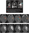
Fig 2. Radiographic appearances of post-LITT changes.
(A) A woman with a left thalamic GBM treated with LITT underwent axial and coronal T1-weighted post-contrast enhanced MR images of the brain pre-LITT, during LITT and 48 hours post LITT. (B-I) A woman with a left insula GBM underwent axial T1-weighted post-contrast enhanced (B-E) and axial FLAIR-weighted (F-I) MR images of the brain pre-LITT and within 48 hours post LITT, 2 weeks post LITT, and 4 weeks post LITT. In both cases, the enhancing tumor (solid red arrowheads in A and B) is replaced by a central zone of T1-weighted signal hyperintensity (open red arrowheads in A and D) and a faint, new discontinuous rim of enhancement extending beyond the original tumor associated enhancing rim (solid blue arrowheads in A and D). The rim of contrast enhancement intensifies at 2 weeks post LITT (D) and remains stable at 4 weeks post LITT (E). Perilesional edema evaluated on FLAIR-weighted images is slightly increased between the pre-treatment (F) and immediate post-treatment (G) images, increases to a maximum point on the 2-week post-treatment images (H) and improves slightly by the 4-week images (I). The orange circles denote a representative ROI used to calculate temporal progression of Ktrans after LITT
Fig 3
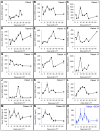
Fig 3. Peritumoral BBB disruption induced by LITT as measured by DCE-MRI.
Ktrans for each of the 14 eligible subjects in the study as a function of time in days from the LITT procedure. In all subjects the Ktrans is highly elevated in the first few days after the procedure and then progressively decreases out to approximately the 4-week time point. This is best illustrated in the bottom right blue graph, which is an average of the 14 subject curves.
Fig 4
Fig 4. Optimization of the BSE ELISA assay for measuring BBB disruption.
Serum concentrations of BSE before and after open craniotomy for surgical debulking in 3 subjects (A, B, and C) with a low-grade glioma, WHO grade II. *p<0.05.
Fig 5
Fig 5. BBB disruption induced by LITT as measured by serum biomarkers.
Serum concentrations of BSE for each of the 14 evaluable subjects in the study (A-N) and as the mean + SEM (O) as a function of time in days from the LITT procedure. In 7/14 subjects, serum BSE levels slightly decreased immediately after LITT, then in 13/14 subjects, serum BSE levels rose shortly after LITT, peaked between 1–3 weeks after LITT, and then decreased by the 6-week time point. In Patient #12, serum BSE concentration increased at week 10 coincident with an increased Ktrans at the same time point, consistent with a recurrent tumor as demonstrated on diagnostic MR imaging. Patient #15’s serum BSE concentration began to rise by week 4, consistent with early multifocal recurrent disease as demonstrated on diagnostic MR imaging.

Comment in

References

    1. Parkin DM. Global cancer statistics in the year 2000. The Lancet Oncology. 2001;2(9):533–43. - PubMed
    1. Stupp R, Mason WP, van den Bent MJ, Weller M, Fisher B, Taphoorn MJB, et al. Radiotherapy plus Concomitant and Adjuvant Temozolomide for Glioblastoma. New England Journal of Medicine. 2005;352(10):987–96. 10.1056/NEJMoa043330 - DOI - PubMed
    1. Stupp R, Hegi ME, Mason WP, van den Bent MJ, Taphoorn MJB, Janzer RC, et al. Effects of radiotherapy with concomitant and adjuvant temozolomide versus radiotherapy alone on survival in glioblastoma in a randomised phase III study: 5-year analysis of the EORTC-NCIC trial. The Lancet Oncology. 2009;10(5):459–66. 10.1016/S1470-2045(09)70025-7 - DOI - PubMed
    1. Hochberg FH, Pruitt A. Assumptions in the radiotherapy of glioblastoma. Neurology. 1980;30(9):907–11. Epub 1980/09/01. . - PubMed
    1. Lemée J-M, Clavreul A, Menei P. Intratumoral heterogeneity in glioblastoma: don't forget the peritumoral brain zone. Neuro-Oncology. 2015;17(10):1322–32. 10.1093/neuonc/nov119 - DOI - PMC - PubMed

Publication types

Associated data