

Development of silica-encapsulated silver nanoparticles as contrast agents intended for dual-energy mammography
- PMID: 26910906
- PMCID: PMC4974128
- DOI: 10.1007/s00330-015-4152-y
Development of silica-encapsulated silver nanoparticles as contrast agents intended for dual-energy mammography
Abstract
Objective: Dual-energy (DE) mammography has recently entered the clinic. Previous theoretical and phantom studies demonstrated that silver provides greater contrast than iodine for this technique. Our objective was to characterize and evaluate in vivo a prototype silver contrast agent ultimately intended for DE mammography.
Methods: The prototype silver contrast agent was synthesized using a three-step process: synthesis of a silver core, silica encapsulation and PEG coating. The nanoparticles were then injected into mice to determine their accumulation in various organs, blood half-life and dual-energy contrast. All animal procedures were approved by the institutional animal care and use committee.
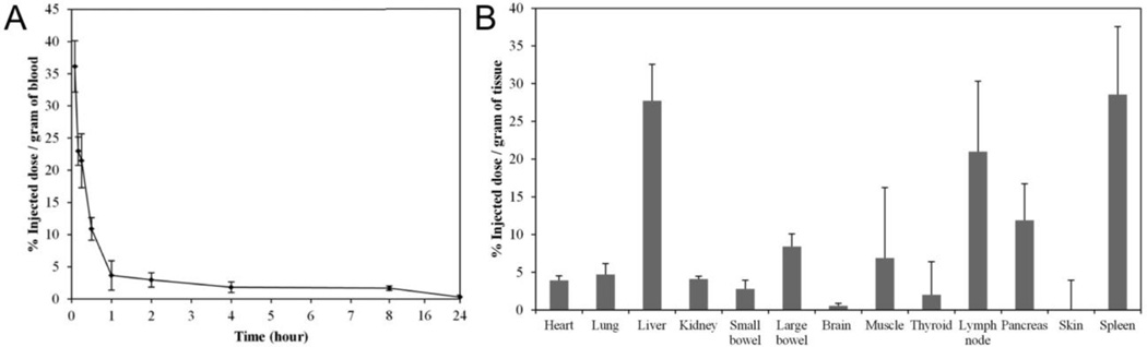
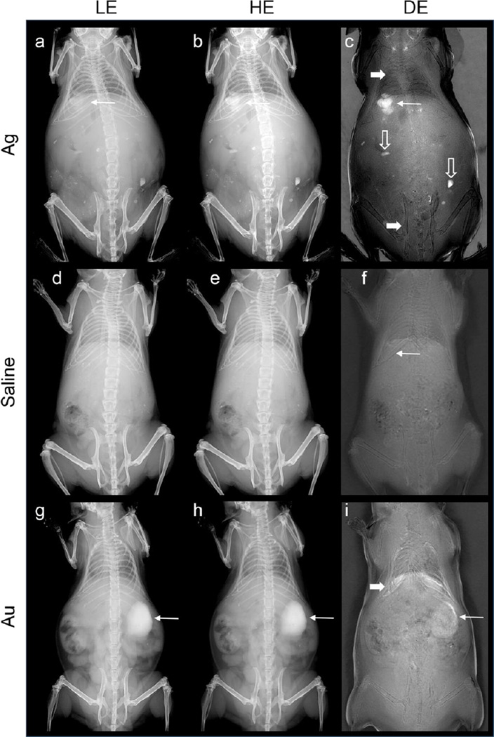
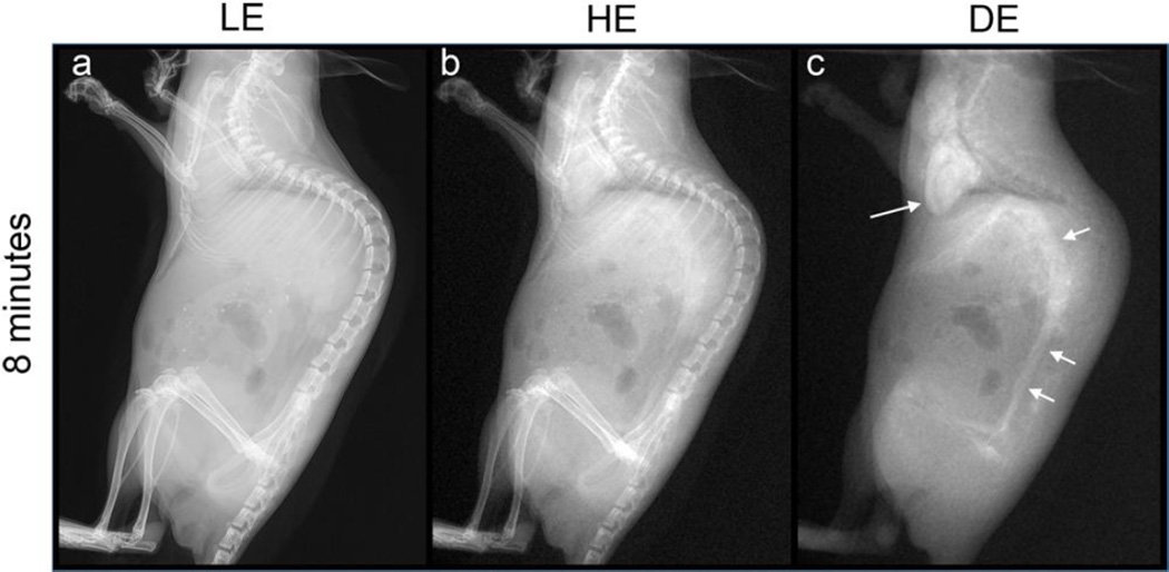
Results: The final diameter of the nanoparticles was measured to be 102 (±9) nm. The particles were removed from the vascular circulation with a half-life of 15 min, and accumulated in macrophage-rich organs such as the liver, spleen and lymph nodes. Dual-energy subtraction techniques increased the signal difference-to-noise ratio of the particles by as much as a factor of 15.2 compared to the single-energy images. These nanoparticles produced no adverse effects in mice.
Conclusion: Silver nanoparticles are an effective contrast agent for dual-energy x-ray imaging. With further design improvements, silver nanoparticles may prove valuable in breast cancer screening and diagnosis.
Key points: • Silver has potential as a contrast agent for DE mammography. • Silica-coated silver nanoparticles are biocompatible and suited for in vivo use. • Silver nanoparticles produce strong contrast in vivo using DE mammography imaging systems.
Keywords: Breast cancer; Dual-energy; Gold; Mammography; Nanoparticles; Silver.
Figures


References
-
- Wang AT, Vachon CM, Brandt KR, K G. Breast density and breast cancer risk: A practical review. Mayo Clin Proc. 2014;89:548–557. - PubMed
-
- Froeling V, Diekmann F, Renz DM, et al. Correlation of contrast agent kinetics between iodinated contrast-enhanced spectral tomosynthesis and gadolinium-enhanced MRI of breast lesions. Eur radiol. 2013;23:1528–1536. - PubMed
Publication types
MeSH terms
Substances
Grants and funding
LinkOut - more resources
Full Text Sources
Other Literature Sources
Medical
