. 2016 Aug;18(8):1169-79.
doi: 10.1093/neuonc/now008. Epub 2016 Feb 23.

Magnetic resonance analysis of malignant transformation in recurrent glioma

Affiliations

Magnetic resonance analysis of malignant transformation in recurrent glioma

Llewellyn E Jalbert et al. Neuro Oncol. 2016 Aug.

Abstract

Background: Patients with low-grade glioma (LGG) have a relatively long survival, and a balance is often struck between treating the tumor and impacting quality of life. While lesions may remain stable for many years, they may also undergo malignant transformation (MT) at the time of recurrence and require more aggressive intervention. Here we report on a state-of-the-art multiparametric MRI study of patients with recurrent LGG.

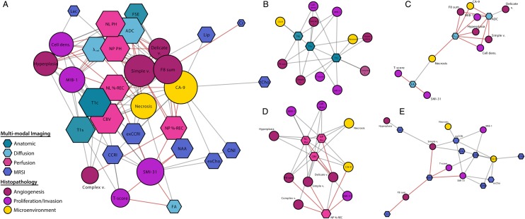
Methods: One hundred and eleven patients previously diagnosed with LGG were scanned at either 1.5 T or 3 T MR at the time of recurrence. Volumetric and intensity parameters were estimated from anatomic, diffusion, perfusion, and metabolic MR data. Direct comparisons of histopathological markers from image-guided tissue samples with metrics derived from the corresponding locations on the in vivo images were made. A bioinformatics approach was applied to visualize and interpret these results, which included imaging heatmaps and network analysis. Multivariate linear-regression modeling was utilized for predicting transformation.

Results: Many advanced imaging parameters were found to be significantly different for patients with tumors that had undergone MT versus those that had not. Imaging metrics calculated at the tissue sample locations highlighted the distinct biological significance of the imaging and the heterogeneity present in recurrent LGG, while multivariate modeling yielded a 76.04% accuracy in predicting MT.

Conclusions: The acquisition and quantitative analysis of such multiparametric MR data may ultimately allow for improved clinical assessment and treatment stratification for patients with recurrent LGG.

Keywords: cancer imaging; low-grade glioma; magnetic resonance; malignant transformation; spectroscopy.


Figures

Fig. 1.
Fig. 1.
Multimodality MRI exam of a subject with a recurrent LGG that had undergone MT. Neurosurgical tissue targets were planned based on regions of suspected tumor using additional functional MR techniques. The imaging revealed a heterogeneously enhancing region of recurrent tumor situated in the left posterior temporal and parietal white matter. An additional, masslike non–contrast enhancing region of residual tumor was also seen in the left insular white matter. This lesion was consistent with residual low-grade neoplasm with marked differences in ADC, PH, and CNI (abnormal regions highlighted in white).
Fig. 2.
Fig. 2.
Heatmaps of volumetric MR parameters and clinical outcome of recurrent LGG. Kaplan–Meier curves generated from (A) OS and (B) PFS demonstrated statistically significant differences among WHO grades for OS between grade II and grade II → III (P = .006), grades II/II → III and GBM (P < .001), while PFS only distinguished GBM from grade II and grade II → III lesions (P < .001). (C) The heatmap generated from patient scans (rows) and hierarchical clustering revealed subgroups of grade II and grade III lesions that displayed abnormal imaging features similar to GBM. Zero volume is shown in blue and approximately twice the median normalized volume is shown in red. Header column denotes the mean parameter value. Gray cells denote cases where there were no data available. NEL, nonenhancing lesion; CEL, contrast enhancing lesion; NEC, necrosis.
Fig. 3.
Fig. 3.
Differences in lesions that had undergone MT. The bar plots represent median differences of volumetric imaging parameter values among the various histological grades and subtypes. Error bars are representative of the 25th and 75th percentiles. Statistical significance was assessed using a Wilcoxon rank sum test at P < .05 (*), P < .01 (**), and P < .005 (***).
Fig. 4.
Fig. 4.
Heatmap of image-guided tissue samples. (A) To assess the biological features within each distinct histology, the neuropathology data were separated by glioma subtype and used hierarchical clustering within each grade. Based on image-guided targeting criteria, several grade II samples were identified that had an unusually high MIB-1 score, and 2 grade III astrocytomas or mixed oligoastrocytomas (OA) had necrosis present. Although few in number, these examples highlight the utility of image guidance to improve tumor sampling. A portion of grade III oligodendroglioma patients had elevated carbonic anhydrase 9 (CA-9) scores of hypoxia. (B) When evaluating the entire mixed population of glioma histologies, we observed a decrease in normal, delicate vasculature (delicate v.), and increases in the presence of simple and complex neovascularization (simple v. and complex v.) were noted within a subset of tumors that had undergone MT.
Fig. 5.
Fig. 5.
Network linkage map of histopathology and advanced imaging of image-guided tissue samples. Nodes were color coded using meta-labels of angiogenesis, proliferation/invasion, and microenvironment for histopathology parameters as well as by modality for imaging parameters. Node size corresponds with average shortest path length. Imaging nodes are presented as hexagons and histopathology nodes as circles. Although imaging and histopathology parameters were intracorrelated, only edges between histopathology and imaging were generated for clarity of network visualization. Red connections denote negative correlation.

Similar articles

Cited by

References

    1. Riemenschneider MJ, Reifenberger G. Molecular neuropathology of low-grade gliomas and its clinical impact. Adv Tech Stand Neurosurg. 2010;35(1):35–64. - PubMed
    1. Grier JT, Batchelor T. Low-grade gliomas in adults. Oncologist. 2006;11(6):681–693. - PubMed
    1. Yan H, Parsons DW, Jin G et al. . IDH1 and IDH2 mutations in gliomas. N Engl J Med. 2009;360(8):765–773. - PMC - PubMed
    1. Houillier C, Wang X, Kaloshi G et al. . IDH1 or IDH2 mutations predict longer survival and response to temozolomide in low-grade gliomas. Neurology. 2010;75(17):1560–1566. - PubMed
    1. Li S, Chou AP, Chen W et al. . Overexpression of isocitrate dehydrogenase mutant proteins renders glioma cells more sensitive to radiation. Neuro Oncol. 2013;15(1):57. - PMC - PubMed

Publication types

MeSH terms