Review
. 2016 Sep;26(9):926-939.
doi: 10.1093/glycob/cww023. Epub 2016 Feb 23.

Plant protein glycosylation

Affiliations
Review

Plant protein glycosylation

Richard Strasser. Glycobiology. 2016 Sep.

Abstract

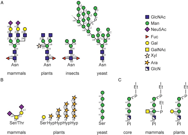
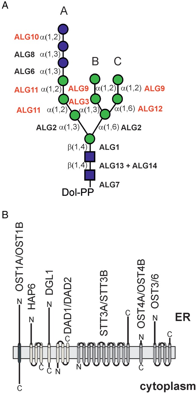
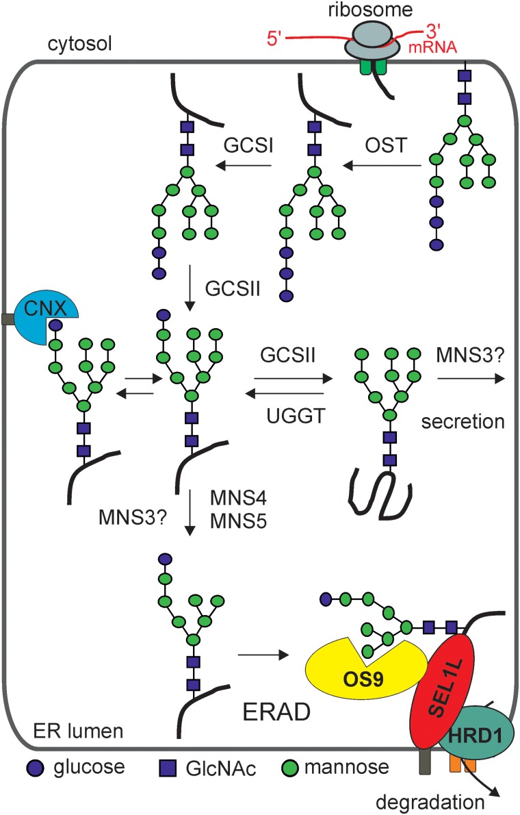
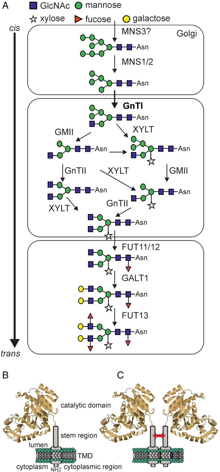
Protein glycosylation is an essential co- and post-translational modification of secretory and membrane proteins in all eukaryotes. The initial steps of N-glycosylation and N-glycan processing are highly conserved between plants, mammals and yeast. In contrast, late N-glycan maturation steps in the Golgi differ significantly in plants giving rise to complex N-glycans with β1,2-linked xylose, core α1,3-linked fucose and Lewis A-type structures. While the essential role of N-glycan modifications on distinct mammalian glycoproteins is already well documented, we have only begun to decipher the biological function of this ubiquitous protein modification in different plant species. In this review, I focus on the biosynthesis and function of different protein N-linked glycans in plants. Special emphasis is given on glycan-mediated quality control processes in the ER and on the biological role of characteristic complex N-glycan structures.

Keywords: Golgi apparatus; N-glycan processing; N-glycosylation; endoplasmic reticulum; glycosyltransferase.


Figures

Fig. 1.
Fig. 1.
Comparison of different types of protein linked glycans. (A) Typical N-glycan structures from mammals, plants, insects and yeast (S. cerevisiae) are shown. The symbols for the monosaccharides in the illustration are drawn according to the nomenclature from the Consortium for Functional Glycomics. (B) Schematic representation of characteristic O-glycans from mammals (di-sialylated core 1), plants (extensin-type modification on Ser and contiguous hydroxyproline (Hyp) residues, the structure is drawn according to Nguema-Ona et al. 2014) and S. cerevisiae. The carbohydrate structure of an A. thaliana arabinogalactan-protein is not shown here and can be found in Tryfona et al. (2012). (C) The conserved core glycan structure of the GPI-anchor is shown as well as examples derived from a human GPI-anchored protein (Kinoshita 2014) and from pear cells (Oxley and Bacic 1999). P-Et indicates the phsphoethanolamine linkage. This figure is available in black and white in print and in color at Glycobiology online.
Fig. 2.
Fig. 2.
(A) ALG proteins involved in lipid-linked oligosaccharide precursor biosynthesis. Genetic evidence for their function has been shown for the A. thaliana glycosyltransferases highlighted in a different color. (A), (B) and (C) define the different branches of the oligosaccharide. (B) Illustration of the putative plant OST complex consisting of different subunits. The proposed topology of the enzymes is depicted. Please note that for some proteins like STT3A/STT3B the exact number of transmembrane domains is unclear (Koiwa et al. 2003) and may range from 10 to 14 helices. The different colors of the transmembrane domains denote subcomplexes that have been described for the yeast and mammalian OST complex (Kelleher and Gilmore 2006). This figure is available in black and white in print and in color at Glycobiology online.
Fig. 3.
Fig. 3.
N-Glycan processing and N-glycan-mediated quality control in the ER of plants. The OST complex transfers the assembled N-glycan precursor to accessible Asn residues within the glycosylation consensus sites of nascent polypeptides. The first N-glycan processing step is carried out by α-glucosidase I (GCSI). Upon trimming of another terminal Glc residue by GCSII, the protein with a mono-glucosylated N-glycan may enter the calnexin (CNX)/calreticulin cycle. Proper folded glycoproteins are released from the quality control process and can exit the ER. Aberrant glycoproteins that cannot attain their final conformation are sent for degradation by the ERAD pathway which requires MNS4/MNS5-mediated Man trimming and recognition by OS9. The class I α-mannosidase MNS3 hydrolyses a single α1,2-Man residue from the middle branch (B-branch see also Figure 2A) of the oligomannosidic N-glycan. MNS3 may act on folded as well as on partially folded glycoproteins. The subcellular site of MNS3 action is still obscure. While MNS3 has so far been exclusively found in Golgi-like structures (Liebminger et al. 2009), ER-resident glycoproteins display N-glycans that have been trimmed by the MNS3-like ER-α-mannosidase activity. This figure is available in black and white in print and in color at Glycobiology online.
Fig. 4.
Fig. 4.
(A) Complex N-glycan formation in the Golgi apparatus. Terminal Man residues are removed by class I α-mannosidases (MNS1-3). Man5GlcNAc2 is used by β1,2-N-acetylglucosaminyltransferase I (GnTI, highlighted in bold) to initiate complex N-glycan formation. The different N-glycan processing enzymes required for the maturation of complex N-glycans in the Golgi are shown. Note, possible cargo transport processes mediated by cisternal maturation, vesicular transport or tubular connections are not indicated. Dependent on the mode of cargo transport there are differences in localization and retention of glycosylation enzymes. (B) Schematic illustration of a Golgi-resident N-glycan processing enzyme. The structure of the catalytic domain from rabbit GnTI (Unligil et al. 2000) is illustrated with N-terminal regions representing the short cytoplasmic tail, the single transmembrane domain and the stem region. (C) Golgi-resident glycosyltransferases may form homomeric or heteromeric complexes that could be important for concentration of the enzymes in different Golgi cisternae and/or for the modulation of their enzymatic activities. The arrow indicates that tobacco GnTI interacts through its stem region (Schoberer et al. 2014). This figure is available in black and white in print and in color at Glycobiology online.

References

    1. Ali MF, Chachadi VB, Petrosyan A, Cheng PW. 2012. Golgi phosphoprotein 3 determines cell binding properties under dynamic flow by controlling Golgi localization of core 2 N-acetylglucosaminyltransferase 1. J Biol Chem. 287:39564–39577. - PMC - PubMed
    1. Atmodjo MA, Sakuragi Y, Zhu X, Burrell AJ, Mohanty SS, Atwood JA, Orlando R, Scheller HV, Mohnen D. 2011. Galacturonosyltransferase (GAUT)1 and GAUT7 are the core of a plant cell wall pectin biosynthetic homogalacturonan:galacturonosyltransferase complex. Proc Natl Acad Sci USA. 108:20225–20230. - PMC - PubMed
    1. Avezov E, Frenkel Z, Ehrlich M, Herscovics A, Lederkremer G. 2008. Endoplasmic reticulum (ER) mannosidase I is compartmentalized and required for N-glycan trimming to Man5-6GlcNAc2 in glycoprotein ER-associated degradation. Mol Biol Cell. 19:216–225. - PMC - PubMed
    1. Bakker H, Bardor M, Molthoff J, Gomord V, Elbers I, Stevens L, Jordi W, Lommen A, Faye L, Lerouge P et al. . 2001. Galactose-extended glycans of antibodies produced by transgenic plants. Proc Natl Acad Sci USA. 98:2899–2904. - PMC - PubMed
    1. Bakker H, Lommen A, Jordi W, Stiekema W, Bosch D. 1999. An Arabidopsis thaliana cDNA complements the N-acetylglucosaminyltransferase I deficiency of CHO Lec1 cells. Biochem Biophys Res Commun. 261:829–832. - PubMed

Publication types