Review
. 2016 May 1;310(10):F931-44.
doi: 10.1152/ajprenal.00523.2015. Epub 2016 Feb 24.

MicroRNAs as novel therapeutic targets to treat kidney injury and fibrosis

Affiliations
Review

MicroRNAs as novel therapeutic targets to treat kidney injury and fibrosis

Ivan G Gomez et al. Am J Physiol Renal Physiol. .

Abstract

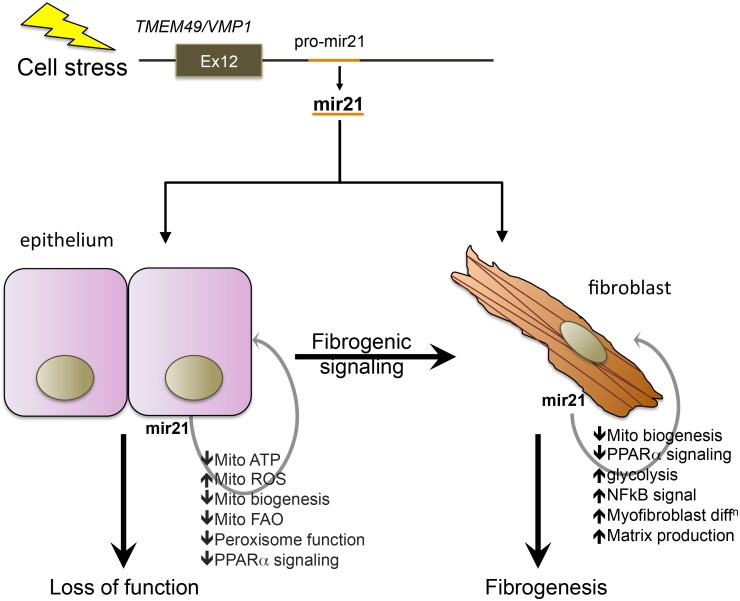
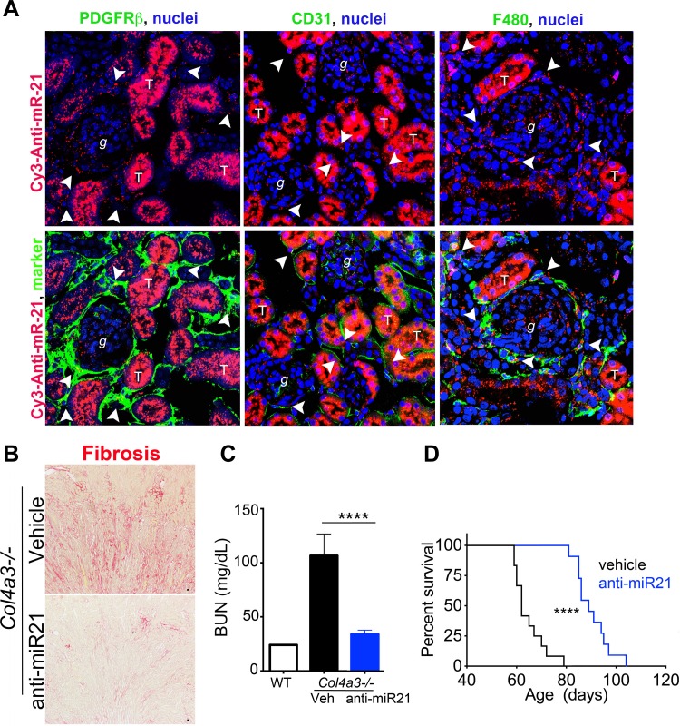
MicroRNAs (miRs), a class of small noncoding RNAs that act as post-transcriptional regulators of gene expression, have attracted increasing attention as critical regulators of organogenesis, cancer, and disease. Interest has been spurred by development of a novel class of synthetic RNA oligonucleotides with excellent drug-like properties that hybridize to a specific miR, preventing its action. In kidney disease, a small number of miRs are dysregulated. These overlap with regulated miRs in nephrogenesis and kidney cancers. Several dysregulated miRs have been identified in fibrotic diseases of other organs, representing a "fibrotic signature," and some of these fibrotic miRs contribute remarkably to the pathogenesis of kidney disease. Chronic kidney disease, affecting ∼10% of the population, leads to kidney failure, with few treatment options. Here, we will explore the pathological mechanism of miR-21, whose pre-eminent role in amplifying kidney disease and fibrosis by suppressing mitochondrial biogenesis and function is established. Evolving roles for miR-214, -199, -200, -155, -29, -223, and -126 in kidney disease will be discussed, and we will demonstrate how studying functions of distinct miRs has led to new mechanistic insights for kidney disease progression. Finally, the utility of anti-miR oligonucleotides as potential novel therapeutics to treat chronic disease will be highlighted.

Keywords: Dicer; angiogenesis; chronic kidney disease; fatty acid oxidation; macrophage activation; microRNAs; mitochondria.


Figures

Fig. 1.
Fig. 1.
Schemas showing synthesis, enzymatic processing, and action of microRNA (miR) as well as the mechanism of miR suppression by anti-miR oligonucleotides. A: miRs are transcribed as 200-nt pri-miRs in the nucleus, cleaved, and processed by Drosha to 70-nt pre-miRs, which are exported to the cytosol where they are assembled into a protein complex with the enzyme Dicer1 which mediates pre-miR processing to the 22-nt mature miR. Mature miRs subsequently assemble with a protein complex (RNA-induced silencing complex; RISC) which includes AGO2, which further cleaves the mature miR to generate a guide from the 3′-arm which contains the seed-matched sequence at the 5′-end of the molecule. The miR associated with the RISC binds to specific sites (seed-matched target sites) at the 3′-end of mRNA, where it facilitates translational repression as well as RNA degradation, thereby inhibiting protein production. B: schema showing the mechanism of action of anti-miR oligonucleotides. Note anti-miRs bind to specific miRs by sequence complementarity in the RISC, thereby preventing anti-miR from interacting with 3′-untranslated regions (UTRs) of target mRNAs. C: image highlighting modifications to the ribose nucleic acid backbone that improve stability, enhance complimentary binding, and enable tissue localization of anti-miR oligonucleotides.
Fig. 2.
Fig. 2.
Identification of regulated miRs in chronic kidney disease. A: graph showing the relative expression of miRs in the human kidney significantly (P < 0.0001) regulated by the presence of chronic kidney disease, defined by albuminuria loss of kidney function and histological fibrosis affecting >25% of the area of the tissue, compared with healthy control kidney biopsies. Clinically indicated biopsies from patients with a kidney transplant and a new kidney disease were collected. Pretransplant biopsies from healthy donors were used as controls. RNA extracted and miR levels were quantified using Agilent arrays and analyzed using Array Studio and DAVID bioinformatics. B: heatmap and Venn diagram showing significantly upregulated miRs comparing a model of obstructive kidney injury [unilateral ureteral obstruction (UUO)] with fibrosis with a model of ischemic injury [ischemia-reperfusion injury (IRI)] with fibrosis. A common signature of 14 overlapping miRs that are upregulated is shown. The heatmap shows relative expression of those upregulated miRs common to both disease models (red is high and blue is low). (Adapted from Ref. 17).
Fig. 3.
Fig. 3.
Schema showing major mechanism of action of miR-21 in accelerating injury responses to promote organ failure and fibrosis. The pre-miR-21 gene is embedded 3′ to the terminal exon of the VMP1 gene, a cell stress response gene involved in autophagy. Upon upregulation, miR-21 directly suppresses genes involved in mitochondrial and peroxisomal functions including biogenesis, fatty acid oxidation, and suppressing reactive oxygen species (ROS) generation. The transcription factor PPARα is an important target for miR-21. In fibroblasts, miR-21, acting partly through PPARα suppression and mitochondrial function suppression, also enhances matrix deposition and NF-κB signaling.
Fig. 4.
Fig. 4.
Graph showing the most significantly enriched gene ontology (GO) pathways in the diseased mouse kidney when miR-21 is absent. KO, knockout.
Fig. 5.
Fig. 5.
Anti-miRNA modified oligonucleotides block specific miRNA function and have excellent drug like properties with high penetrance to kidney cells in vivo. (A) Fluorescence images (X400) of the kidney showing the distribution of a single injection of anti-miR21-Cy3 (red) subcutaneously (25 mg/kg in 50 μl, 48 h previously). Note that the compound is concentrated in tubule epithelium but is also detected in glomerular cells, macrophages (F4/80), endothelium (CD31) and fibroblasts (PDGFRβ). Arrowheads show areas of co-localization. Effect of Anti-miR21 on (B) Fibrosis, (C) Blood urea nitrogen concentration (D) and survival (I) in Col4a3−/− mice with Alport nephropathy. (Adapted from Ref. 37). ****P < 0.0001.

References

    1. Ahmed MI, Alam M, Emelianov VU, Poterlowicz K, Patel A, Sharov AA, Mardaryev AN, Botchkareva NV. MicroRNA-214 controls skin and hair follicle development by modulating the activity of the Wnt pathway. J Cell Biol 207: 549–567, 2014. - PMC - PubMed
    1. Alvarez ML, Khosroheidari M, Eddy E, Kiefer J. Role of microRNA 1207-5P and its host gene, the long non-coding RNA Pvt1, as mediators of extracellular matrix accumulation in the kidney: implications for diabetic nephropathy. PLoS One 8: e77468, 2013. - PMC - PubMed
    1. Androsavich JR, Chau BN, Bhat B, Linsley PS, Walter NG. Disease-linked microRNA-21 exhibits drastically reduced mRNA binding and silencing activity in healthy mouse liver. RNA 18: 1510–1526, 2012. - PMC - PubMed
    1. Anglicheau D, Sharma VK, Ding R, Hummel A, Snopkowski C, Dadhania D, Seshan SV, Suthanthiran M. MicroRNA expression profiles predictive of human renal allograft status. Proc Natl Acad Sci USA 106: 5330–5335, 2009. - PMC - PubMed
    1. Azzouzi el H, Leptidis S, Dirkx E, Hoeks J, van Bree B, Brand K, McClellan EA, Poels E, Sluimer JC, van den Hoogenhof MMG, Armand AS, Yin X, Langley S, Bourajjaj M, Olieslagers S, Krishnan J, Vooijs M, Kurihara H, Stubbs A, Pinto YM, Krek W, Mayr M, da Costa Martins PA, Schrauwen P, De Windt LJ. The hypoxia-inducible microRNA cluster miR-199a∼214 targets myocardial PPARδ and impairs mitochondrial fatty acid oxidation. Cell Metab 18: 341–354, 2013. - PubMed

Publication types

LinkOut - more resources