Review
. 2016 Apr;7(2):245-54.
doi: 10.1007/s13244-016-0470-0. Epub 2016 Feb 25.

MRI evaluation prior to Transcatheter Aortic Valve Implantation (TAVI): When to acquire and how to interpret

Affiliations
Review

MRI evaluation prior to Transcatheter Aortic Valve Implantation (TAVI): When to acquire and how to interpret

Abhishek Chaturvedi et al. Insights Imaging. 2016 Apr.

Abstract

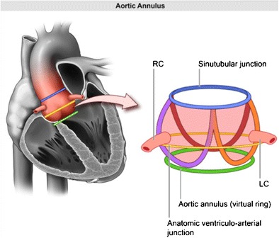
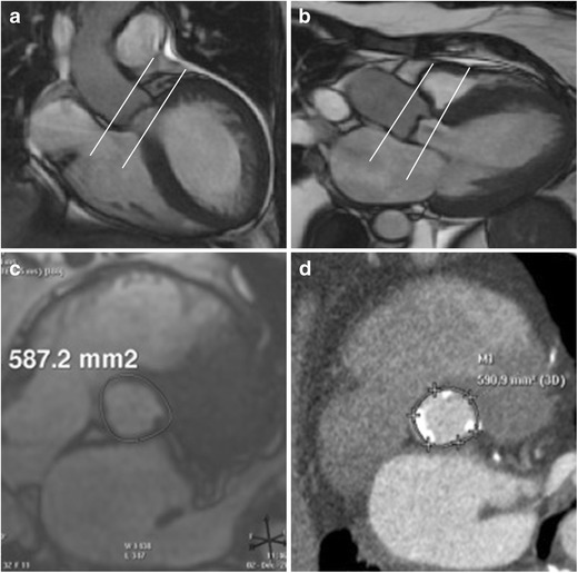
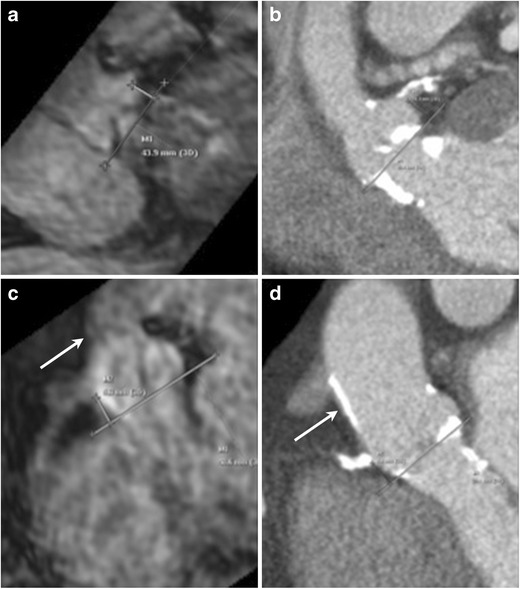
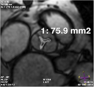
Transcatheter Aortic Valve Implantation (TAVI) is increasingly being used in patients with severe aortic stenosis who are not candidates for surgery. ECG-gated CT angiography (CTA) plays an important role in the preoperative planning for these devices. As the number of patients undergoing these procedures increases, a subset of patients is being recognized who have contraindications to iodinated contrast medium, either due to a prior severe allergic type reaction or poor renal function. Another subgroup of patients with low flow and low gradient aortic stenosis is being recognized that are usually assessed for severity of aortic stenosis by stress echocardiography. There are contraindications to stress echocardiography and some of these patients may not be able to undergo this test. Non-contrast MRI can be a useful emerging modality for evaluating these patients. In this article, we discuss the emerging indications of non-contrast MRI in preoperative assessment for TAVI and describe the commonly used MRI sequences. A comparison of the most important measurements obtained for TAVI assessment on CTA and MRI from same subjects is included. Teaching Points • MRI can be used for preoperative assessment of aortic annulus. • MRI is an alternate to CTA when iodinated contrast is contraindicated. • Measurements obtained by non-contrast MRI are similar to contrast enhanced CTA. • MRI can be used to assess severity of aortic stenosis.

Keywords: Magnetic resonance imaging; Non-contrast MRI; Severe aortic stenosis; Transcatheter aortic valve implantation; Transcatheter aortic valve replacement.


Figures

Fig. 1
Fig. 1
Pictorial depiction of the aortic root complex demonstrating the location of the annulus, aortoannular, ventriculoarterial and sinotubular junction
Fig. 2
Fig. 2
Acquisition on MRI for aortic annular plane to measure diameters, area, and perimeter. Aortic root cine stack is prescribed from coronal aorta (a) and left ventricle outflow tract views (b). Systolic image where the luminal diameter is widest, in a location just below the insertion of the valve leaflets (c) is identified as the annular slice. Corresponding annular image from the same patient obtained from ECG-gated CTA demonstrates similar measurement of annular area (d)
Fig. 3
Fig. 3
Assessment of coronary ostial height on MRI. Navigator-assisted 3-D SSFP stack of the aortic root in the late diastolic phase is acquired. Ostial height is measured from the aortic valve annular plane (a: left coronary artery, c right coronary artery). In the same patient corresponding CT images (b: left coronary artery, d right coronary artery). This MRI sequence can be also be used to assess sinus of valsalva height and width. Note the dark appearance of the anterior aortic wall and the left ventricle outflow tract due to extensive calcifications, easily seen on the corresponding CTA images
Fig. 4
Fig. 4
Assessment of aortic stenosis by planimetry. 2-D cine SSFP acquisition parallel to the valve plane demonstrates the narrowest opening of the aortic orifice. CT of the same patient also demonstrated similar orifice area
Fig. 5
Fig. 5
Diameters of sinuses of valsalva measured on MRI. 2-D SSFP of the aortic root in late diastolic phase is identified from the aortic root stack. Diameters are measured in mid-sinus above the aortic valve (a: noncoronary, b: left and c right). In the same patient corresponding CT images (d: noncoronary, e: left coronary and f: right coronary). Navigator-assisted 3-D SSFP or T2 black blood sequences can also be used for these measurements
Fig. 6
Fig. 6
Identification of access site in tubular ascending aortic for transaortic implantation on MRI. Skin to aortic distance measured on axial non-gated SSFP sequence (a) similar to CTA (b). Access site on aorta to annular plane distance measured from multiplanar sagittal oblique reformats obtained from the same axial non-gated SSFP sequence (c). Same measurement in this patient obtained from CTA (d)
Fig. 7
Fig. 7
Assessment of left ventricle apex on MRI. Location of the apex in the intercostal space and the distance of this access site from the midline can be measured and marked using axial non-gated SSFP sequence (a). Same site location by CTA in this patient (b)

References

    1. Bennett CJ, Maleszewski JJ, Araoz PA. CT and MR imaging of the aortic valve: radiologic-pathologic correlation. Radiographics. 2012;32:1399–1420. doi: 10.1148/rg.325115727. - DOI - PubMed
    1. Leon MB, Smith CR, Mack M, et al. Transcatheter aortic-valve implantation for aortic stenosis in patients who cannot undergo surgery. N Engl J Med. 2010;363:1597–1607. doi: 10.1056/NEJMoa1008232. - DOI - PubMed
    1. Quail MA, Nordmeyer J, Schievano S, Reinthaler M, Mullen MJ, Taylor AM. Use of cardiovascular magnetic resonance imaging for TAVR assessment in patients with bioprosthetic aortic valves: comparison with computed tomography. Eur J Radiol. 2012;81:3912–3917. doi: 10.1016/j.ejrad.2012.07.014. - DOI - PubMed
    1. (2013) ACR Manual on contrast media
    1. Pontone G, Andreini D, Bartorelli AL, et al. Aortic annulus area assessment by multidetector computed tomography for predicting paravalvular regurgitation in patients undergoing balloon-expandable transcatheter aortic valve implantation: a comparison with transthoracic and transesophageal echocardiography. Am Heart J. 2012;164:576–584. doi: 10.1016/j.ahj.2012.06.024. - DOI - PubMed

LinkOut - more resources