. 2016 Feb 24:47:34.
doi: 10.1186/s13567-016-0319-z.

Japanese encephalitis virus tropism in experimentally infected pigs

Affiliations

Japanese encephalitis virus tropism in experimentally infected pigs

Meret E Ricklin et al. Vet Res. .

Abstract

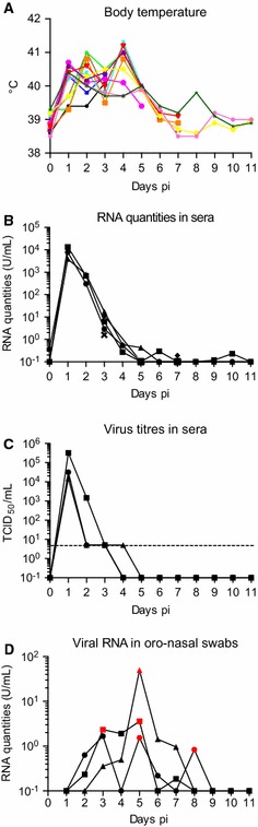
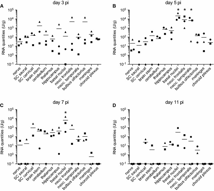
Pigs are considered to be the main amplifying host for Japanese encephalitis virus (JEV), and their infection can correlate with human cases of disease. Despite their importance in the ecology of the virus as it relates to human cases of encephalitis, the pathogenesis of JEV in pigs remains obscure. In the present study, the localization and kinetics of virus replication were investigated in various tissues after experimental intravenous infection of pigs. The data demonstrate a rapid and broad spreading of the virus to the central nervous system (CNS) and various other organs. A particular tropism of JEV in pigs not only to the CNS but also for secondary lymphoid tissue, in particular the tonsils with the overall highest viral loads, was observed. In this organ, even 11 days post infection, the latest time point of the experiment, no apparent decrease in viral RNA loads and live virus was found despite the presence of a neutralizing antibody response. This was also well beyond the clinical and viremic phase. These results are of significance for the pathogenesis of JEV, and call for further experimental studies focusing on the cellular source and duration of virus replication in pigs.


Figures

Figure 1
Figure 1
Development of body temperature and viremia following experimental infection of pigs with JEV. 12 piglets were infected with 107 TCID50 JEV Nakayama strain. Pigs were killed in groups of three on days 3, 5, 7, and 11 pi. From three pigs daily serum samples were obtained, and from nine pigs blood samples were collected at necropsy (days 3, 5, 7 and 11 pi). A Daily body temperature of all animals. Each colour represents an individual animal. B Viral RNA loads in the serum. Relative JEV RNA quantities were determined using real-time RT-PCR. Quantification employed a standard curve created using a viral preparation with a known infectious titre. One U was defined as the viral RNA quantity corresponding to 1 TCID50 of the virus preparation used as standard. C Viral titres in the serum of individual bled daily. The detection limit of the assay was 5 TCID50/mL (dotted line). D Viral RNA loads in the oro-nasal swabs collected daily from three different animals. The red symbols represent samples from which live virus isolation succeeded.
Figure 2
Figure 2
Viral RNA loads in the peripheral organs following experimental infection of pigs with JEV. At necropsy peripheral organs were sampled and the tissue homogenized before quantitative RT-PCR was run. Relative RNA quantities are shown for each day (AD) calculated in U/mL as explained in the legend of Figure 1 and Materials and methods. Bars indicate mean values of a group. To illustrate RT-PCR negative samples they were plotted as 10−1 U/mL. *difference statistically significant (p < 0.05) when this organ is compared to all others for a particular time point pi.
Figure 3
Figure 3
Viral RNA loads in the nervous system following experimental infection of pigs with JEV. At necropsy nervous tissues were sampled and homogenized before quantitative RT-PCR was run. Relative RNA quantities are shown for each day (AD) calculated in U/mL. Bars indicate mean values of a group. To illustrate RT-PCR negative samples they were plotted as 10−1 U/mL. *difference statistically significant (p < 0.05) when this organ is compared to all others for a particular time point pi.
Figure 4
Figure 4
Neuropathology in the olfactory bulb of a JEV-infected pig at 5 days pi (score 3). A Histopathology of the olfactory bulb (bub) and tract (tract). Multifocal glial nodules were present in the grey matter of the olfactory bulb (arrows) and accompanied by mononuclear perivascular cuffs (arrowheads). H and E, 20× magnification, bar = 500 µm. B Higher magnification (400×) of one glial nodule shown in A). One degenerated, shrunken neuron (arrow) in the olfactory bulb is surrounded by a nodule of microglial and inflammatory cells. H and E, bar = 20 µm.
Figure 5
Figure 5
Histological scores in the CNS following JEV-infection of pigs. At necropsy brains and the spinal cord were sampled, fixed and processed routinely for H and E staining. Data are shown for each day (AD) separately. Lesions were scored semi-quantitatively from 0 to 4 (0=no lesions, 1=minimal lesions, 2=mild lesions, 3=moderate lesions, 4=severe lesions). Scores of three healthy control piglets (CTRL) were 0. E Sum of all CNS tissues scores with each symbol representing an individual pig.
Figure 6
Figure 6
Development of neutralizing antibodies following experimental infection of pigs with JEV. All sera were tested for their neutralization ability using a standard plaque neutralization assay. Titres are calculated as the last serum dilution that reduces viral plaques by 50%.

References

    1. Impoinvil DE, Baylis M, Solomon T. Japanese encephalitis: on the one health agenda. Curr Top Microbiol Immunol. 2013;365:205–247. - PubMed
    1. van den Hurk AF, Ritchie SA, Mackenzie JS. Ecology and geographical expansion of Japanese encephalitis virus. Annu Rev Entomol. 2009;54:17–35. doi: 10.1146/annurev.ento.54.110807.090510. - DOI - PubMed
    1. Campbell GL, Hills SL, Fischer M, Jacobson JA, Hoke CH, Hombach JM, Marfin AA, Solomon T, Tsai TF, Tsu VD, Ginsburg AS. Estimated global incidence of Japanese encephalitis: a systematic review. Bull World Health Organ. 2011;89:766–774. doi: 10.2471/BLT.10.085233. - DOI - PMC - PubMed
    1. Weaver SC, Barrett AD. Transmission cycles, host range, evolution and emergence of arboviral disease. Nat Rev Microbiol. 2004;2:789–801. doi: 10.1038/nrmicro1006. - DOI - PMC - PubMed
    1. Endy TP, Nisalak A. Japanese encephalitis virus: ecology and epidemiology. Curr Top Microbiol Immunol. 2002;267:11–48. - PubMed

Publication types