Review
. 2016 Apr:52:84-92.
doi: 10.1016/j.semcdb.2016.02.004. Epub 2016 Feb 18.

Metabolic remodeling in early development and cardiomyocyte maturation

Affiliations
Review

Metabolic remodeling in early development and cardiomyocyte maturation

Rebecca Ellen Kreipke et al. Semin Cell Dev Biol. 2016 Apr.

Abstract

Aberrations in metabolism contribute to a large number of diseases, such as diabetes, obesity, cancer, and cardiovascular diseases, that have a substantial impact on the mortality rates and quality of life worldwide. However, the mechanisms leading to these changes in metabolic state--and whether they are conserved between diseases--is not well understood. Changes in metabolism similar to those seen in pathological conditions are observed during normal development in a number of different cell types. This provides hope that understanding the mechanism of these metabolic switches in normal development may provide useful insight in correcting them in pathological cases. Here, we focus on the metabolic remodeling observed both in early stage embryonic stem cells and during the maturation of cardiomyocytes.

Keywords: Cardiac maturation; Cardiomyocyte; Developmental transitions; Metabolism; Naïve to primed embryonic stem cell transition; Pluripotent stem cells; hESC.


Figures

Figure 1
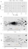
Figure 1. Differential expression of genes between primed and naïve human embryonic stem cell lines
(a) Principal component analysis (PCA) of RNA-seq data from published primed and naïve hESC lines, using all genes [22]. Each dot represents a sample. Different primed and naïve lines are color- and shape-coded. Primed hESC lines form a cluster on the left, naïve hESC lines form a cluster on the right. (b) Genes contributing to principal components separating primed versus naïve hESCs. Each dot represents a gene. The size of the dots is proportional to the contribution of the gene to the primed versus naïve separation along PC1 (x-axis in a,[22]). Top contributing genes are darker. (c) Metabolic genes contributing to principal components separating primed versus naïve hESCs when only metabolic genes are used in the PCA analysis. The size of the dots is proportional to the contribution of the gene to the primed versus naïve separation along PC1 (x-axis). Top contributing genes are darker. Genes in b and c are listed in supplemental tables 1 and 2.
Figure 2
Figure 2. Differential expression of genes between fetal and adult cardiomyocytes
(a) Principal component analysis (PCA) of RNA-seq data from published human fetal and adult heart samples, as well as 20 day and 1 year old cardiomyocytes, using all genes. Each dot represents a sample. (b) Genes contributing to principal components separating fetal (fetal, 20 day, and 1 year old) versus adult heart samples. Each dot represents a gene. The size of the dots is proportional to the contribution of the gene to the fetal versus adult separation along PC1 (x-axis in a). Top contributing genes are darker. (c) Metabolic genes contributing to principal components separating fetal (fetal, day 20, and 1 year old) versus adult heart sample when only metabolic genes are used in the PCA analysis. The size of the dots is proportional to the contribution of the gene to the fetal versus adult separation along PC1 (x-axis). Top contributing genes are darker. Genes in b and c are listed in supplemental tables 3 and 4.

References

    1. Folmes CDL, Nelson TJ, Martinez-Fernandez A, Arrell DK, Lindor JZ, Dzeja PP, et al. Somatic oxidative bioenergetics transitions into pluripotency-dependent glycolysis to facilitate nuclear reprogramming. Cell Metab. 2011;14:264–271. - PMC - PubMed
    1. Panopoulos AD, Yanes O, Ruiz S, Kida YS, Diep D, Tautenhahn R, et al. The metabolome of induced pluripotent stem cells reveals metabolic changes occurring in somatic cell reprogramming. Cell Res. 2012;22:168–177. - PMC - PubMed
    1. Yanes O, Clark J, Wong DM, Patti GJ, Sánchez-Ruiz A, Benton HP, et al. Metabolic oxidation regulates embryonic stem cell differentiation. Nat Chem Biol. 2010;6:411–417. - PMC - PubMed
    1. Bracha AL, Ramanathan A, Huang S, Ingber DE, Schreiber SL. Carbon metabolism–mediated myogenic differentiation. Nat Chem Biol. 2010;6:202–204. - PMC - PubMed
    1. Greer SN, Metcalf JL, Wang Y, Ohh M. The updated biology of hypoxia-inducible factor. EMBO J. 2012;31:2448–2460. - PMC - PubMed