. 2016 Feb 24:9:13.
doi: 10.1186/s13045-016-0244-7.

H2S protects against fatal myelosuppression by promoting the generation of megakaryocytes/platelets

Affiliations

H2S protects against fatal myelosuppression by promoting the generation of megakaryocytes/platelets

Huan-Di Liu et al. J Hematol Oncol. .

Abstract

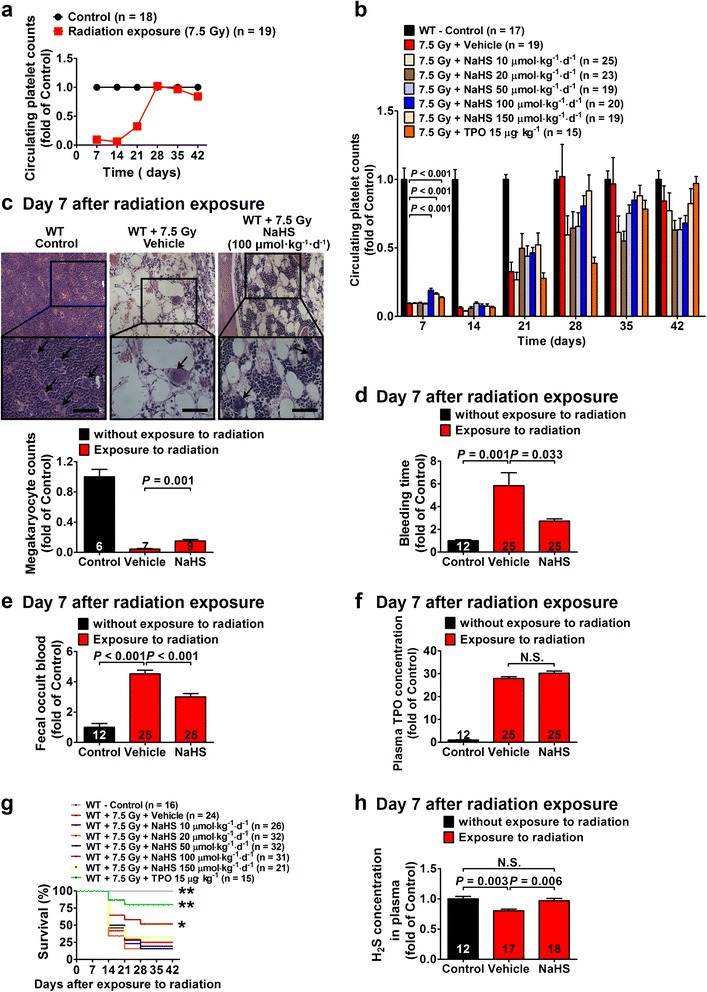
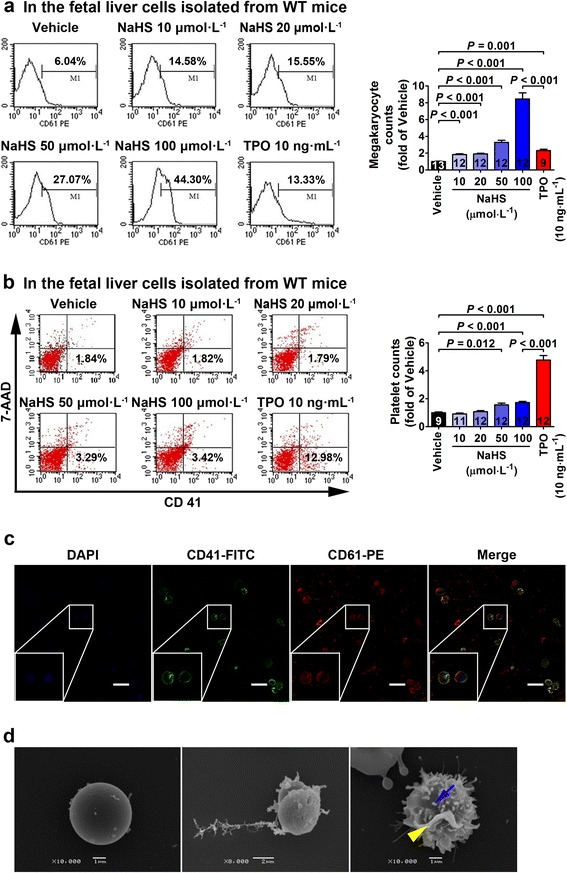
Background: Our previous pilot studies aimed to examine the role of hydrogen sulfide (H2S) in the generation of endothelial progenitor cells led to an unexpected result, i.e., H2S promoted the differentiation of certain hematopoietic stem/progenitor cells in the bone marrow. This gave rise to an idea that H2S might promote hematopoiesis.

Methods: To test this idea, a mice model of myelosuppression and cultured fetal liver cells were used to examine the role of H2S in hematopoiesis.

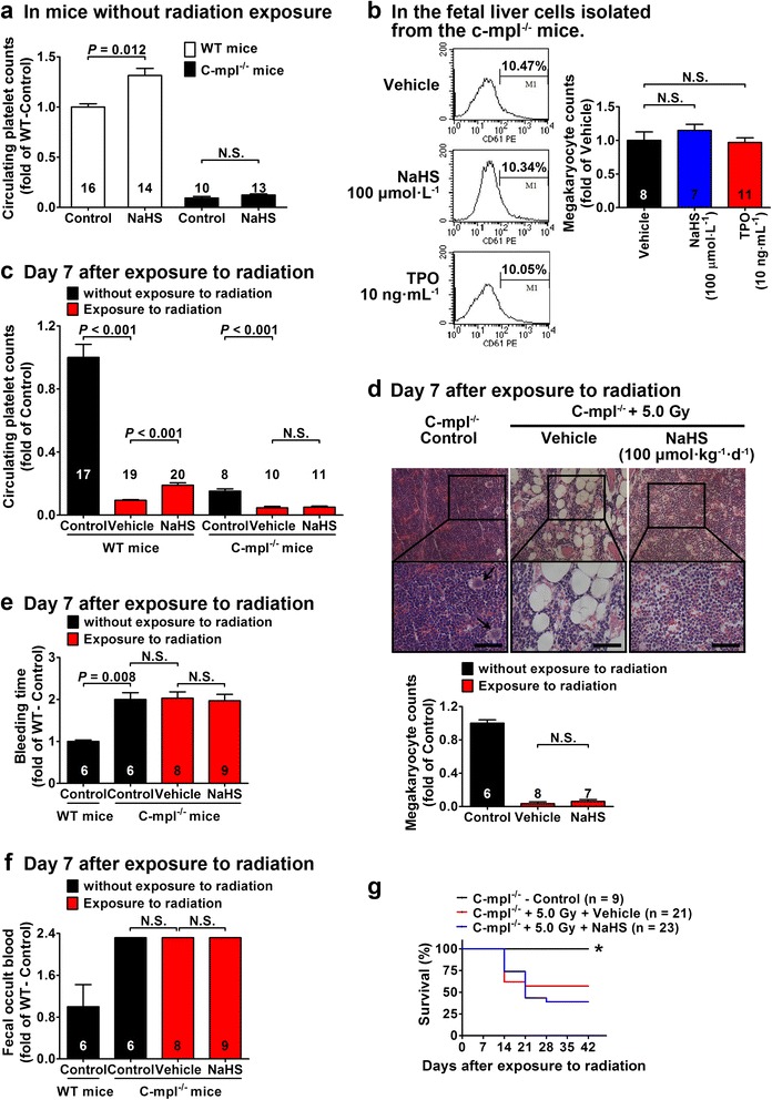
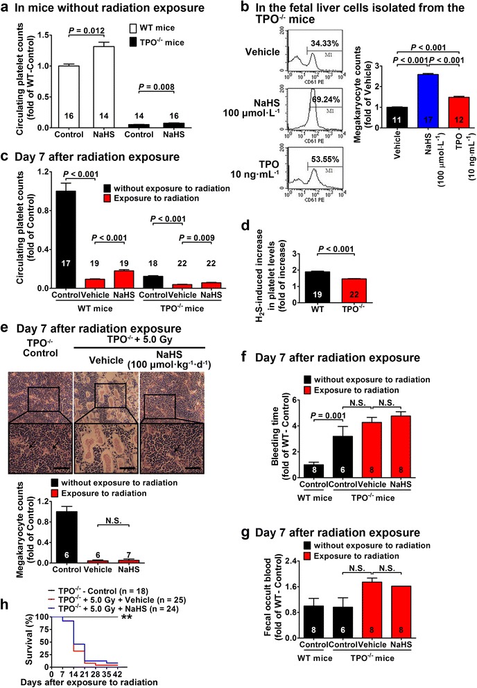
Results: H2S promoted the generation of megakaryocytes, increased platelet levels, ameliorate entorrhagia, and improved survival. These H2S effects were blocked in both in vivo and in vitro models with thrombopoietin (TPO) receptor knockout mice (c-mpl(-/-) mice). In contrast, H2S promoted megakaryocytes/platelets generation in both in vivo and in vitro models with TPO knockout mice (TPO(-/-) mice).

Conclusions: H2S is a novel promoter for megakaryopoiesis by acting on the TPO receptors but not TPO to generate megakaryocytes/platelets.


Figures

Fig. 1
Fig. 1
H2S treatment promotes platelet generation, alleviates bleeding, and improves survival in WT mice with radiation-induced myelosuppression. a Time course of circulating platelet counts after exposure to radiation of 137Cs at a single dose of 7.5 Gy. b NaHS treatment caused a significant increase in circulating platelet levels at 100 and 150 μmol kg−1 day−1 on day 7 after radiation exposure. TPO treatment also provided a similar amelioration at 15 μg kg−1. c Morphological examination showed a significant decrease in the number of megakaryocytes (black arrow) in the bone marrow of WT mice exposed to radiation, whereas NaHS (100 μmol kg−1 day−1) significantly increased the number of megakaryocytes as compared with the vehicle-treated group. Scale bar = 250 μm. d Bleeding time was prolonged after exposure to radiation, and bleeding was ameliorated by NaHS treatment (100 μmol kg−1 day−1) as compared with that of vehicle-treated WT mice. e Likewise, fecal occult blood was significantly increased after exposure to radiation and NaHS treatment (100 μmol kg−1 day−1) caused a significant decrease in fecal occult blood in mice exposed to radiation. f Plasma TPO concentrations were increased in WT mice on day 7 after radiation exposure where NaHS treatment (100 μmol kg−1 day−1) did not change TPO concentrations. g Radiation exposure resulted in a significant decrease in survival as compared with that of the mice without radiation exposure. Both NaHS (100 μmol kg−1 day−1) and TPO (15 μg kg−1) treatment significantly improved survival in these mice exposed to radiation. *P < 0.05, **P < 0.01 versus vehicle-treated mice exposed to radiation, log-rank test. h Plasma H2S levels were decreased in mice exposed to radiation, and this decrease was recovered by administration of NaHS (100 μmol kg−1 day−1). Data in the graphs are means ± SEM. P values less than 0.05 represent statistical significance
Fig. 2
Fig. 2
H2S treatment promotes generation of megakaryocytes/platelets in cultured fetal liver cells. a NaHS treatment caused a concentration-dependent increase in megakaryocytes which were identified as CD61-positive cells using flow cytometry in fetal liver cells isolated from WT mice. b Platelets were identified as CD41-positive and 7-AAD-negative signals using flow cytometry. Platelet counts were significantly increased with NaHS treatment at 50 and 100 μmol L−1 in the culture medium of fetal liver cells. TPO treatment (10 ng mL−1) also increased platelet counts. c Morphology of megakaryocytes in cultured fetal liver cells were shown with a confocal fluorescence microscopy with staining of DAPI, CD41-FITC, and CD61-PE. Scale bar = 50 μm. d Scanning electron microscopy revealed pseudopod formation (blue arrow) and membrane blebbing (yellow arrow) in the megakaryocytes identified in cultured fetal liver cells treated with NaHS (100 μmol L−1). Data in the graphs are means ± SEM. P values less than 0.05 represent statistical significance
Fig. 3
Fig. 3
H2S-induced promotion of megakaryocytes/platelets generation and improvement of survival are blunted in c-mpl−/− mice. a Without radiation exposure, NaHS treatment (20 μmol kg−1 day−1) for 14 days caused an increase in circulating platelet levels in WT mice but not in c-mpl−/− mice. b Flow cytometry showed that NaHS treatment (100 μmol L−1) failed to increase the number of megakaryocytes identified as CD61-positive cells in cultured fetal liver cells isolated from c-mpl−/− mice. c Circulating platelets were significantly decreased in both WT and c-mpl−/− mice exposed to radiation at day 7. NaHS treatment (100 μmol kg−1 day−1) increased platelet levels in WT mice but not in c-mpl−/− mice. d Morphological examination of the bone marrow showed that NaHS treatment (100 μmol kg−1 day−1) failed to increase the number of megakaryocytes (black marrows) in c-mpl−/− mice. Scale bar = 250 μm. e Bleeding was not ameliorated with NaHS treatment (100 μmol kg−1 day−1) in c-mpl−/− mice exposed to radiation at day 7. f Likewise, fecal occult blood was not ameliorated with NaHS treatment (100 μmol kg−1 day−1) in c-mpl−/− mice exposed to radiation at day 7. g NaHS treatment (100 μmol kg−1 day−1) failed to improve survival in c-mpl−/− mice exposed to radiation as compared to vehicle-treated mice. *P < 0.05 versus vehicle-treated mice exposed to radiation, log-rank test. Data in the graphs are means ± SEM. P values less than 0.05 represent statistical significance
Fig. 4
Fig. 4
H2S promotes megakaryocytes/platelets generation but does not improve survival in TPO−/− mice. a Without radiation exposure, NaHS treatment (20 μmol kg−1 day−1) for 14 days caused an increase in circulating platelet levels in both WT and TPO−/− mice. b NaHS treatment (100 μmol L−1) increased the number of megakaryocytes identified as CD61-positive cells using flow cytometry in cultured fetal liver cells isolated from TPO−/− mice. c, d NaHS treatment (100 μmol kg−1 day−1) increased circulating platelet levels on day 7 after radiation exposure in both WT and TPO−/− mice (c). However, the effects of NaHS treatment (100 μmol kg−1 day−1) in increasing circulating platelet levels were blunted in TPO−/−mice (d). e Morphological examination of the bone marrow showed that NaHS treatment (100 μmol kg−1 day−1) did not increase the number of megakaryocytes (black arrows) in the TPO−/− mice exposed to radiation. Scale bar = 250 μm. f Bleeding was not ameliorated with NaHS treatment (100 μmol kg−1 day−1) in the TPO−/− mice exposed to radiation at day 7. g Likewise, fecal occult blood was not ameliorated with NaHS treatment (100 μmol kg−1 day−1) in the TPO−/− mice exposed to radiation at day 7. h Survival was not improved with NaHS treatment (100 μmol kg−1 day−1) in TPO−/− mice exposed to radiation. **P < 0.01 versus vehicle-treated mice exposed to radiation, log-rank test. Data in the graphs are means ± SEM. P values less than 0.05 represent statistical significance

References

    1. Henry MK, Lynch JT, Eapen AK, Quelle FW. DNA damage-induced cell-cycle arrest of hematopoietic cells is overridden by activation of the PI-3 kinase/Akt signaling pathway. Blood. 2001;98(3):834–41. doi: 10.1182/blood.V98.3.834. - DOI - PubMed
    1. Mauch P, Constine L, Greenberger J, et al. Hematopoietic stem cell compartment: acute and late effects of radiation therapy and chemotherapy. Int J Radiat Oncol Biol Phys. 1995;31(5):1319–39. doi: 10.1016/0360-3016(94)00430-S. - DOI - PubMed
    1. Wang Y, Schulte BA, LaRue AC, Ogawa M, Zhou D. Total body irradiation selectively induces murine hematopoietic stem cell senescence. Blood. 2006;107(1):358–66. doi: 10.1182/blood-2005-04-1418. - DOI - PMC - PubMed
    1. Li J, Yang C, Xia Y, et al. Thrombocytopenia caused by the development of antibodies to thrombopoietin. Blood. 2001;98(12):3241–8. doi: 10.1182/blood.V98.12.3241. - DOI - PubMed
    1. Schrezenmeier H, Seifried E. Buffy-coat-derived pooled platelet concentrates and apheresis platelet concentrates: which product type should be preferred? Vox Sang. 2010;99(1):1–15. doi: 10.1111/j.1423-0410.2009.01295.x. - DOI - PubMed

Publication types

MeSH terms

LinkOut - more resources