. 2016 Feb 25:6:22086.
doi: 10.1038/srep22086.

8-Oxoguanine accumulation in mitochondrial DNA causes mitochondrial dysfunction and impairs neuritogenesis in cultured adult mouse cortical neurons under oxidative conditions

Affiliations

8-Oxoguanine accumulation in mitochondrial DNA causes mitochondrial dysfunction and impairs neuritogenesis in cultured adult mouse cortical neurons under oxidative conditions

Julio Leon et al. Sci Rep. .

Erratum in

Abstract

Oxidative stress and mitochondrial dysfunction are implicated in aging-related neurodegenerative disorders. 8-Oxoguanine (8-oxoG), a common oxidised base lesion, is often highly accumulated in brains from patients with neurodegenerative disorders. MTH1 hydrolyses 8-oxo-2'-deoxyguanosine triphosphate (8-oxo-dGTP) to 8-oxo-dGMP and pyrophosphate in nucleotide pools, while OGG1 excises 8-oxoG paired with cytosine in DNA, thereby minimising the accumulation of 8-oxoG in DNA. Mth1/Ogg1-double knockout (TO-DKO) mice are highly susceptible to neurodegeneration under oxidative conditions and show increased accumulation of 8-oxoG in mitochondrial DNA (mtDNA) in neurons, suggesting that 8-oxoG accumulation in mtDNA causes mitochondrial dysfunction. Here, we evaluated the contribution of MTH1 and OGG1 to the prevention of mitochondrial dysfunction during neuritogenesis in vitro. We isolated cortical neurons from adult wild-type and TO-DKO mice and maintained them with or without antioxidants for 2 to 5 days and then examined neuritogenesis. In the presence of antioxidants, both TO-DKO and wild-type neurons exhibited efficient neurite extension and arborisation. However, in the absence of antioxidants, the accumulation of 8-oxoG in mtDNA of TO-DKO neurons was increased resulting in mitochondrial dysfunction. Cells also exhibited poor neurite outgrowth with decreased complexity of neuritic arborisation, indicating that MTH1 and OGG1 are essential for neuritogenesis under oxidative conditions.


Figures

Figure 1
Figure 1. Localisation of MTH1 and OGG1 proteins in adult mouse cerebral cortex.
(a) Genotyping of wild-type and Mth1/Ogg1-double knockout (TO-DKO) mice. Wild-type (Mth1+, Ogg1+) and mutant (Mth1, Ogg1) alleles were amplified using specific primer sets. (b) Cortical tissues from wild-type and TO-DKO male mice were subjected to western blotting with antibodies against MTH1, OGG1, and β-actin as a loading control. (c) Immunohistochemical detection of MTH1 in wild-type neocortex, layers 1–3 (left panel) and layers 4–5 (right panel). (d) Immunohistochemical detection of OGG1 in wild-type neocortex, layers 1–3 (left panel) and layers 4–5 (right panel). Scale bar = 50 μm.
Figure 2
Figure 2. Cortical neurons isolated from adult wild-type and Mth1/Ogg1-DKO brains regenerate neurites.
(a) Purity of cortical neuron culture at day 3 in vitro. Neuronal (MAP2, green) and microglial (F4/80, red) markers were detected by immunofluorescence microscopy. Nuclei were counter stained by DAPI (blue). About 95% of the cultured cells were MAP2-positive neurons. Merged images are shown. Scale bar = 50 μm. (b) Phase contrast images of cortical neurons isolated from adult wild-type (left panels) and Mth1/Ogg1-DKO (TO-DKO) (right panels) mice cultured for 0, 3 and 6 days, showing time-dependent neurite regeneration. Scale bar = 20 μm.
Figure 3
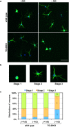
Figure 3. Neuritogenesis in cortical neurons isolated from adult Mth1/Ogg1-DKO but not wild-type mice was significantly impaired in the absence of antioxidants.
(a) Adult cortical neurons isolated from Mth1/Ogg1-DKO (TO-DKO) and wild-type mice were cultured for 2 days in the absence (−AO) or presence (+AO) of antioxidants in B27 supplements, and were subjected to MAP2-immunofluorescence microscopy. Green: MAP2, blue: DAPI. Representative merged images are shown. Scale bar = 50 μm. (b) Regenerating neurons were classified into three stages: stage 1, lacking neurites; stage 2, with one or more minor neurite; stage 3, with one neurite at least twice as long as any other. Representative images of neurons in each stage are shown. (c) Distribution of regenerating neurons. Percentage of neurons in each stage is shown. Error bar = SEM. N = 4 independent experiments. More than 50 neurons in each culture condition were examined. *Fisher’s exact test: p < 0.0001 vs. other sample in each stage.
Figure 4
Figure 4. Neurite arborisation of cortical neurons isolated from adult Mth1/Ogg1-DKO but not wild-type mice was significantly impaired in the absence of antioxidants.
(a) Adult cortical neurons isolated from Mth1/Ogg1-DKO (TO-DKO) and wild-type mice were cultured for 5 days in the absence (−AO) or presence (+AO) of antioxidants and were subjected to MAP2-immunofluorescence microscopy. Representative images are shown, indicating a simpler neuritic branching pattern in neurons from TO-DKO mice cultured in the absence of antioxidants. Scale bar = 20 μm. (b) Sholl’s concentric sphere analysis shows severely impaired neurite arborisation in TO-DKO neurons in the absence of antioxidants. More than 16 neurons were examined in each culture condition. Combined data from two independent experiments are shown. Error bar = SEM. MANOVA, between subjects: F(3, 171) = 19.671, p < 0.0001; within subjects: interaction, p < 0.0001 (Univar G-G, ε = 0.311). TO-DKO (−AO) vs. other three groups, *p < 0.0001.
Figure 5
Figure 5. Mth1/Ogg1-DKO neurons do not exhibit increased cell death in the absence of antioxidants.
(a) Adult cortical neurons isolated from adult Mth1/Ogg1-DKO (TO-DKO) and wild-type mice were cultured for 2 days in vitro (2DIV, left panels) and 5 days in vitro (5DIV, right panels) in the absence (−AO) or presence (+AO) of antioxidants in the B27 supplements, and were subjected to Hoechst 33258 (blue, nuclear staining) and propidium iodide (PI, red, dead cells) staining. Representative images are shown. (b) Viability. The percentage of PI-negative cells among Hoechst 33258-positive cells was determined. 2DIV (left, 2-way ANOVA, F(3, 16) = 0.7804, p = 0.522), and 5DIV (right, 2-way ANOVA, F(3, 16) = 0.1565, p = 0.924). Error bar = SEM. More than 11 cells in a given field were counted and five different fields per group (>79 cells/group) were examined. Scale bar = 40 μm.
Figure 6
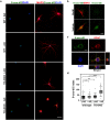
Figure 6. MTH1/OGG1 deficiency significantly increased accumulation of 8-oxoguanine in the mitochondrial DNA of cortical neurons in the absence of antioxidants.
(a) 8-Oxo-deoxyguanosine (8-oxo-dG) detected in MAP2-positive neurons by immunofluorescence microscopy. Fixed neurons pre-treated with RNase were subjected to a mild denaturation with 25 mM NaOH before reacting with antibodies. Adult cortical neurons isolated from Mth1/Ogg1-DKO (TO-DKO) and wild-type (WT) mice were cultured for 2 days in the absence (−AO) or presence (+AO) of antioxidants. Green: 8-oxo-dG; red: MAP2: blue: DAPI. Scale bar = 20 μm. Cytoplasmic 8-oxo-dG immunoreactivity was increased in TO-DKO neurons maintained in the absence of antioxidant. (b) 8-Oxo-dG immunoreactivity in TO-DKO neurons in the absence of antioxidants was completely abolished by pre-treatment with MutM 8-oxoG DNA glycosylase. Scale bar = 10 μm. (c) Mitochondrial localization of 8-oxo-dG in a TO-DKO neuron. Immunofluorescence signals for mitochondrial voltage-dependent anion channel (VDAC, red) were co-localized with the cytoplasmic 8-oxo-dG immunofluorescence (green). Orthogonal views obtained by laser scanning confocal microscopy are shown. Blue: DAPI. Scale bar = 10 μm. (d) Quantitative evaluation of mitochondrial 8-oxo-dG in adult cortical neurons with (+AO) or without (−AO) antioxidants. More than 203 cells were examined for each group. 8-Oxo-dG indexes were calculated and are presented as whisker-box plots. Outliers are shown as dots. Wilcoxon/Kruskal–Wallis tests, chi square test p < 0.0001; post hoc comparison with Wilcoxon method, *p < 0.05, **p < 0.005, ***p < 0.0001.
Figure 7
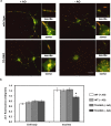
Figure 7. MTH1/OGG1 deficiencies cause mitochondrial dysfunction in adult cortical neurons.
(a) Adult cortical neurons isolated from Mth1/Ogg1-DKO (TO-DKO) and wild-type brains were cultured for 2 days in the absence (−AO) or presence (+AO) of antioxidants and were then exposed to JC-1 dye. JC-1 dye emits green-fluorescence in its monomer form with lower membrane potential, and red-fluorescence in its aggregate form in normal mitochondria. Scale bar = 20 μm. (b) Red/green ratio of JC-1 fluorescence in cell body (soma in a) and neurites. Mitochondrial membrane potentials were significantly decreased in TO-DKO neurons cultured without antioxidants. 2-way ANOVA, F(3, 80) = 7.495, p = 0.0002 *Tukey-HSD test, TO-DKO (−AO) vs. other three groups. p < 0.002. More than 17 neurons were examined for each condition. Error bar = SEM.
Figure 8
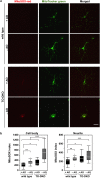
Figure 8. MTH1/OGG1-defciencies significantly increased mitochondrial production of superoxide in adult cortical neurons cultured in the absence of antioxidants.
(a) Adult cortical neurons isolated from Mth1/Ogg1-DKO (TO-DKO) and wild-type brains were cultured for 2 days in the absence (−AO) or presence (+AO) of antioxidants, then incubated with MitoTracker (green) and MitoSOX (red). Representative images are shown. Scale bar = 20 μm. (b) MitoSOX index in cell body and neurites are shown as whisker-box plots. Mitochondrial superoxide production significantly increased in TO-DKO neurons cultured in the absence of antioxidants. More than 17 neurons were examined for each condition. Outliers are shown as dots. Wilcoxon/Kruskal–Wallis tests, chi square test p < 0.0001; post hoc comparison with Wilcoxon method, *p < 0.05, **p < 0.005, ***p < 0.0001.

Similar articles

Cited by

References

    1. Riemer J. & Kins S. Axonal transport and mitochondrial dysfunction in Alzheimer’s disease. Neurodegener Dis 12, 111–124 (2013). - PubMed
    1. Hokama M. et al. Altered expression of diabetes-related genes in Alzheimer’s disease brains: the hisayama study. Cereb Cortex 24, 2476–2488 (2014). - PMC - PubMed
    1. Henchcliffe C. & Beal M. F. Mitochondrial biology and oxidative stress in Parkinson disease pathogenesis. Nat Clin Pract Neurol 4, 600–609 (2008). - PubMed
    1. Ayala-Pena S. Role of oxidative DNA damage in mitochondrial dysfunction and Huntington’s disease pathogenesis. Free Radic Biol Med 62, 102–110 (2013). - PMC - PubMed
    1. Kang D., Takeshige K., Sekiguchi M. & Singh K. K. Introduction. In: Mitochondrial DNA mutations in aging, disease and cancer (ed. Singh K. K. ) Ch. 1, 1–15 (Springer-Verlag, 1999).

Publication types