

Evaluation of serum and tissue levels of VAP-1 in colorectal cancer
- PMID: 26912327
- PMCID: PMC4766640
- DOI: 10.1186/s12885-016-2183-7
Evaluation of serum and tissue levels of VAP-1 in colorectal cancer
Abstract
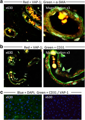
Background: The endothelial adhesion molecule, vascular adhesion protein-1 (VAP-1, AOC3) promotes lymphocyte recruitment to tumours, although the contribution that VAP-1 makes to lymphocyte recruitment in human colorectal cancer (CRC) is unknown. VAP-1 exists in circulating soluble form (sVAP-1). A previous study demonstrated elevated sVAP-1 levels in CRC patients. The aim of this study was to confirm this finding and study the differences in tissue VAP-1 expression between CRC and healthy tissues.
Methods: sVAP-1 levels were measured in the serum of 31 patients with CRC and 31 age- and sex-matched controls. Tissue VAP-1 levels were measured by immunohistochemistry, quantitative real-time PCR and Western blotting.
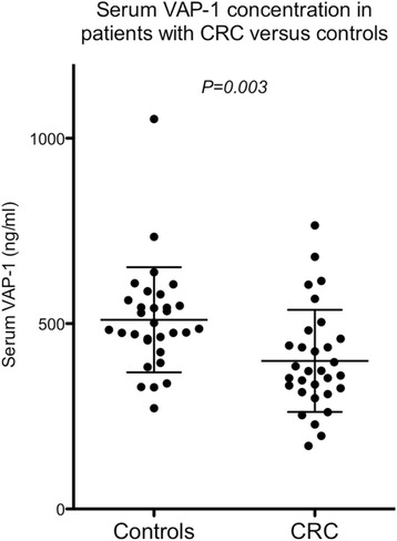
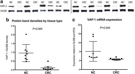
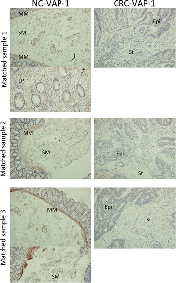
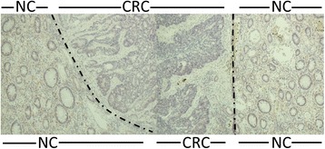
Results: The mean sVAP-1 level ± SD was significantly lower in the CRC group compared with the control group (399 ± 138 ng/ml versus 510 ± 142 ng/ml, P = 0.003). Tissue VAP-1 protein and mRNA levels were significantly lower in CRC compared with normal colon tissue. VAP-1 immunostaining was practically absent from CRC.
Conclusions: VAP-1 is downregulated in human CRC and although the molecular basis of this down regulation is not yet known, we suggest it may be part of a mechanism used by the tumour to prevent the recruitment of anti-tumour immune cells. Our data contradicts the findings of others with regard sVAP-1 levels in patients with CRC. Possible reasons for this are discussed.
Figures
References
-
- Cancer Incidence and Mortality in the United Kingdom, 2008-10 [http://www.ons.gov.uk/ons/rel/cancer-unit/cancer-incidence-and-mortality...]
-
- Edwards BK, Noone A-M, Mariotto AB, Simard EP, Boscoe FP, Henley SJ, Jemal A, Cho H, Anderson RN, Kohler BA, Eheman CR, Ward EM. Annual Report to the Nation on the status of cancer, 1975-2010, featuring prevalence of comorbidity and impact on survival among persons with lung, colorectal, breast, or prostate cancer. Cancer 2013:n/a–n/a. - PMC - PubMed
-
- Mlecnik B, Tosolini M, Kirilovsky A, Berger A, Bindea G, Meatchi T, Bruneval P, Trajanoski Z, Fridman W-H, Pagès F, Galon J. Histopathologic-Based Prognostic Factors of Colorectal Cancers Are Associated With the State of the Local Immune Reaction. J Clin Oncol. 2011;29:610–618. doi: 10.1200/JCO.2010.30.5425. - DOI - PubMed
Publication types
MeSH terms
Substances
Grants and funding
LinkOut - more resources
Full Text Sources
Other Literature Sources
Medical
Research Materials
