. 2016 Mar 11;351(6278):1214-8.
doi: 10.1126/science.aad5214. Epub 2016 Feb 11.

MTAP deletion confers enhanced dependency on the PRMT5 arginine methyltransferase in cancer cells

Affiliations

MTAP deletion confers enhanced dependency on the PRMT5 arginine methyltransferase in cancer cells

Gregory V Kryukov et al. Science. .

Abstract

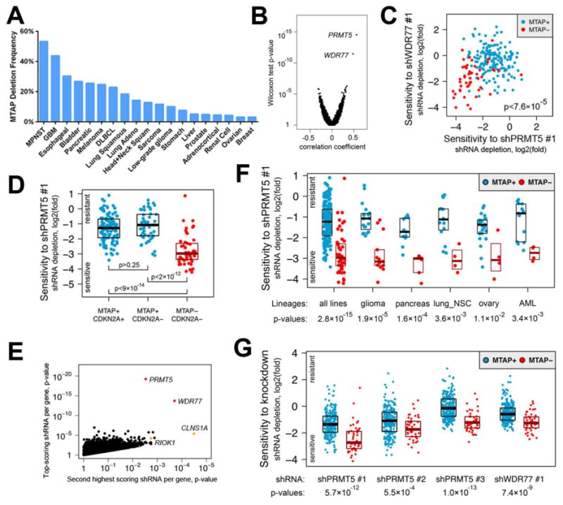
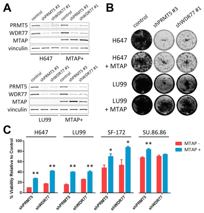
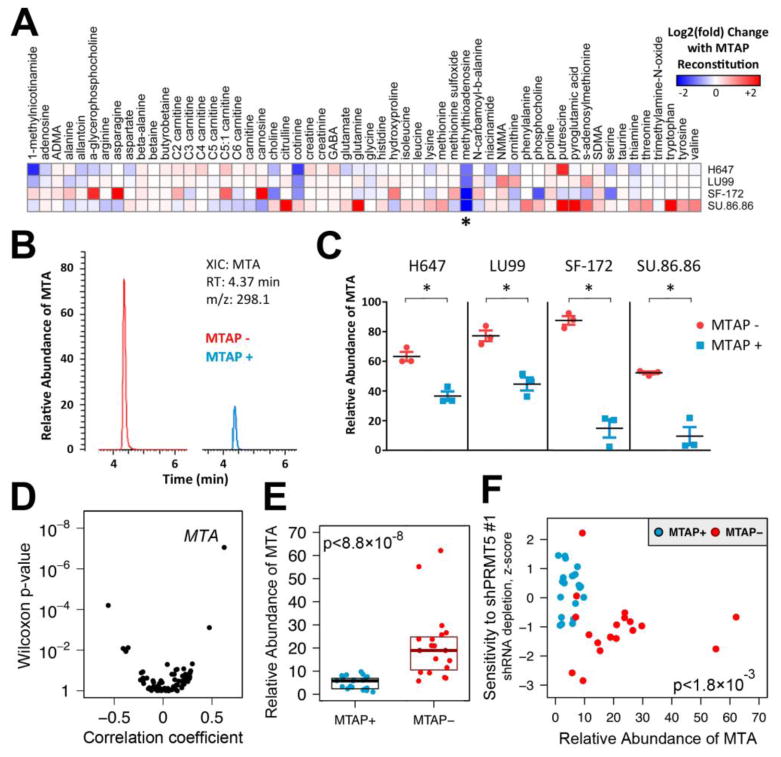
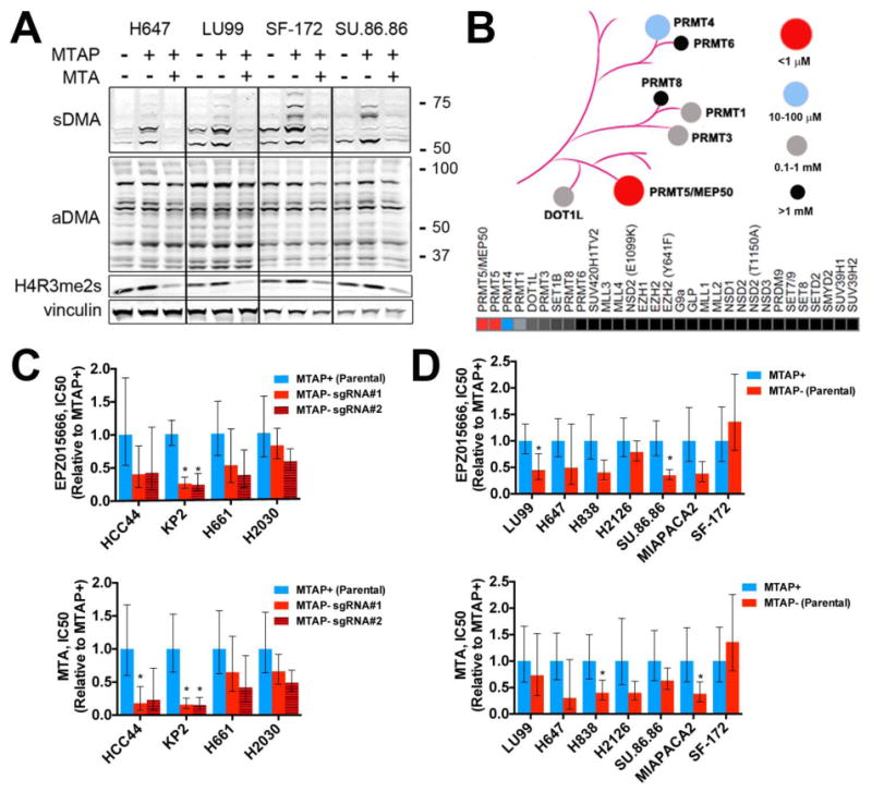
The discovery of cancer dependencies has the potential to inform therapeutic strategies and to identify putative drug targets. Integrating data from comprehensive genomic profiling of cancer cell lines and from functional characterization of cancer cell dependencies, we discovered that loss of the enzyme methylthioadenosine phosphorylase (MTAP) confers a selective dependence on protein arginine methyltransferase 5 (PRMT5) and its binding partner WDR77. MTAP is frequently lost due to its proximity to the commonly deleted tumor suppressor gene, CDKN2A. We observed increased intracellular concentrations of methylthioadenosine (MTA, the metabolite cleaved by MTAP) in cells harboring MTAP deletions. Furthermore, MTA specifically inhibited PRMT5 enzymatic activity. Administration of either MTA or a small-molecule PRMT5 inhibitor showed a modest preferential impairment of cell viability for MTAP-null cancer cell lines compared with isogenic MTAP-expressing counterparts. Together, our findings reveal PRMT5 as a potential vulnerability across multiple cancer lineages augmented by a common "passenger" genomic alteration.


Figures

Fig. 1
Fig. 1. Cancer cell lines with homozygous MTAP loss are selectively sensitive to suppression of PRMT5 or WDR77
(A) Frequency of MTAP deletion for selected cancers is shown. Data was obtained from the cBioPortal for Cancer Genomics (http://www.cbioportal.org). MPNST, malignant peripheral nerve sheath tumor; GBM, glioblastoma; DLBCL, diffuse large B-cell lymphoma. (B) Point biserial correlation coefficients are plotted against Wilcoxon two-class comparison test p-values for 50,529 shRNAs. (C) Log2(fold) of depletion of shPRMT5 #1 and shWDR77 #1 are shown, demonstrating a correlation between sensitivity to these shRNAs for MTAP− lines. (D) Log2(fold) of shPRMT5 #1 depletion is plotted for cell lines with the indicated genotypes. Median with upper and lower 25th percentiles are shown. (E) Pearson correlation test p-values for the top-scoring shRNAs are plotted against p-values for the second best-scoring shRNAs targeting the same gene. Selective sensitivity of MTAP− lines to depletion of the methylosome is supported by at least two hairpins against four members of the complex including constitutive members of the complex (PRMT5 and WDR77, red) and mutually exclusive substrate adaptors (CLNS1A and RIOK1, orange). (F) Log2(fold) of shPRMT5 #1 depletion is plotted for all 216 cell lines (left) and for lines from the indicated lineages. lung_NSC, non-small cell lung cancer; AML, acute myeloid leukemia. (G) Log2(fold) depletion for the indicated shRNAs is shown for all 275 cell lines from the validation cohort.
Fig. 2
Fig. 2. Cells with MTAP loss are more sensitive to suppression of PRMT5 and WDR77 than isogenic MTAP-reconstituted cells
(A) Protein lysates were harvested from H647 (top) or LU99 (bottom) and from MTAP-reconstituted H647 or LU99 cells (MTAP+) 5 days after lentiviral transduction with the indicated shRNAs or control. Lysates were fractionated by SDS-PAGE, and immunoblotting was performed with the indicated antibodies. (B) H647 or LU99 cells and MTAP-reconstituted H647 or LU99 cells were transduced with lentivirus harboring the indicated shRNAs and stained with crystal violet after 10 to 18 days. Media change was performed every 3 days. (C) Quantitation of crystal violet uptake by cells transduced with shRNAs against PRMT5 or WDR77 (normalized to control shRNA for each cell line). Mean and standard error of 3-4 replicates are shown. The experiment was performed 2-3 times for each of the four cell line pairs. ** p < 0.01; * p < 0.05 by two-tailed Student's t-test.
Fig. 3
Fig. 3. Intracellular MTA is increased in MTAP− cells and correlates with sensitivity to PRMT5 suppression
(A) Relative abundance of 56 profiled metabolites was compared for cell extracts from 4 isogenic cell line pairs. Fold-change in relative abundance of each metabolite with MTAP reconstitution is shown for each isogenic pair. Results represent the mean of 2 independent experiments with 3 replicates per cell line. Findings for MTA (methylthioadenosine) are indicated with an asterisk. (B) Representative extracted ion chromatograms (XICs) from LC-MS analysis of SF-172 (left) or MTAP-reconstituted SF-172 (right) cell extracts demonstrating a peak corresponding to MTA. RT, retention time; m/z, mass-to-charge ratio. (C) Relative abundance of MTA from cell extracts is displayed. Mean and standard error of 3 biological replicates are shown. The experiment was performed twice with similar findings. * p < 0.01 by Student's t-test. (D) Correlation of metabolite levels with MTAP status is shown. Point biserial correlation coefficients are plotted against Wilcoxon two-class comparison test p-values for 73 metabolites profiled across 40 MTAP+ and MTAP−cell lines. (E) Relative abundance of MTA from MTAP+ (n=21) and MTAP− (n=19) cell lines from various lineages is shown. For each cell line, mean of 3 biological replicates is displayed. Median with upper and lower 25th percentiles are shown for MTAP− and MTAP+ lines. (F) Correlation of intracellular MTA levels with sensitivity to PRMT5 depletion is shown. shPRMT5 sensitivity data from the screening and validation studies was normalized and combined using modified z-scores. Z-scores are plotted against relative intracellular abundance of MTA for the 40 assayed cell lines. Spearman rank correlation p-value is shown.
Fig. 4
Fig. 4. Pharmacological inhibition of PRMT5
(A) Cells were exposed to DMSO or 200 μM MTA for 48 hours. Lysates were harvested and fractionated by SDS-PAGE. Immunoblotting was performed with the indicated antibodies [recognizing symmetric or asymmetric di-methyl arginine motifs (sDMA and aDMA, respectively), symmetric di-methyl histone H4 arginine 3 (H4R3me2s), or vinculin (loading control)]. Molecular weight is indicated on the right in kDa. (B) Dendrogram and heat map indicating relative sensitivity of 31 histone methyltransferases to inhibition by MTA as determined by radioisotope filter binding assay. (C, D) Relative cell viability IC50 (normalized to MTAP+ cells for each line) for cells treated with EPZ015666 (top) or MTA (bottom), for isogenic cell lines derived from (C) MTAP-expressing parental cell lines or (D) MTAP− parental cell lines. Mean IC50 and 95% confidence interval of 6 replicates are shown for each cell line. Each experiment was performed twice for each cell line. * = p < 0.05 by two-tailed Student's t-test (vs. MTAP+ cells).

Comment in

References

    1. Nobori T, Takabayashi K, Tran P, Orvis L, Batova A, Yu AL, Carson DA. Genomic cloning of methylthioadenosine phosphorylase: a purine metabolic enzyme deficient in multiple different cancers. Proc Natl Acad Sci U S A. 1996;93:6203–6208. - PMC - PubMed
    1. Chen ZH, Zhang H, Savarese TM. Gene deletion chemoselectivity: codeletion of the genes for p16(INK4), methylthioadenosine phosphorylase, and the alpha- and beta-interferons in human pancreatic cell carcinoma lines and its implications for chemotherapy. Cancer Res. 1996;56:1083–1090. - PubMed
    1. Illei PB, Rusch VW, Zakowski MF, Ladanyi M. Homozygous deletion of CDKN2A and codeletion of the methylthioadenosine phosphorylase gene in the majority of pleural mesotheliomas. Clin Cancer Res. 2003;9:2108–2113. - PubMed
    1. Hustinx SR, Hruban RH, Leoni LM, Iacobuzio-Donahue C, Cameron JL, Yeo CJ, Brown PN, Argani P, Ashfaq R, Fukushima N, Goggins M, Kern SE, Maitra A. Homozygous deletion of the MTAP gene in invasive adenocarcinoma of the pancreas and in periampullary cancer: a potential new target for therapy. Cancer Biol Ther. 2005;4:83–86. - PubMed
    1. Powell EL, Leoni LM, Canto MI, Forastiere AA, Iocobuzio-Donahue CA, Wang JS, Maitra A, Montgomery E. Concordant loss of MTAP and p16/CDKN2A expression in gastroesophageal carcinogenesis: evidence of homozygous deletion in esophageal noninvasive precursor lesions and therapeutic implications. Am J Surg Pathol. 2005;29:1497–1504. - PubMed

Publication types

MeSH terms