. 2016 Mar 15;113(11):E1506-15.
doi: 10.1073/pnas.1601569113. Epub 2016 Feb 24.

FOXC1 maintains the hair follicle stem cell niche and governs stem cell quiescence to preserve long-term tissue-regenerating potential

Affiliations

FOXC1 maintains the hair follicle stem cell niche and governs stem cell quiescence to preserve long-term tissue-regenerating potential

Kenneth Lay et al. Proc Natl Acad Sci U S A. .

Abstract

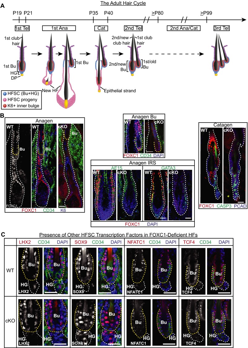
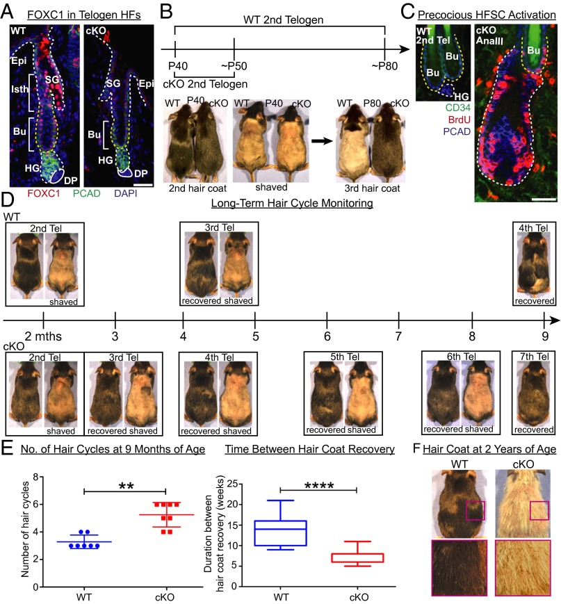
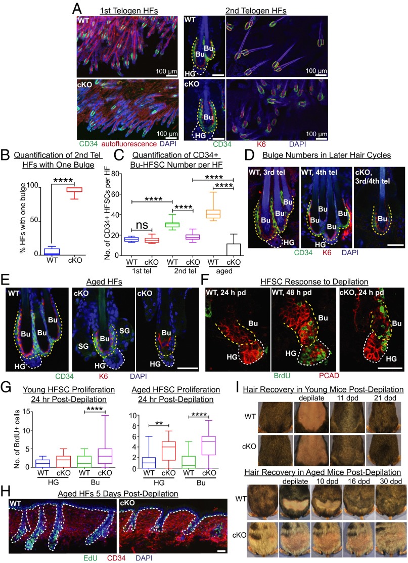
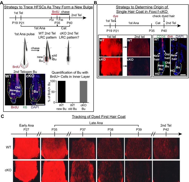
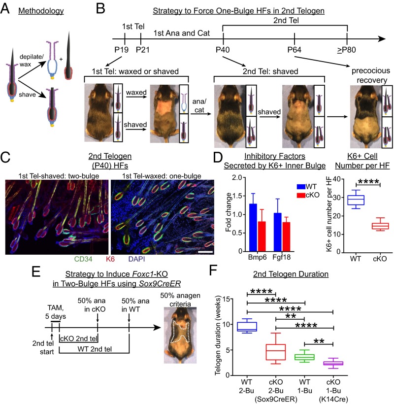
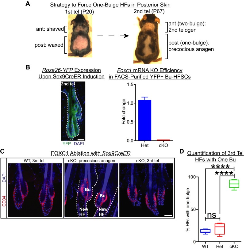
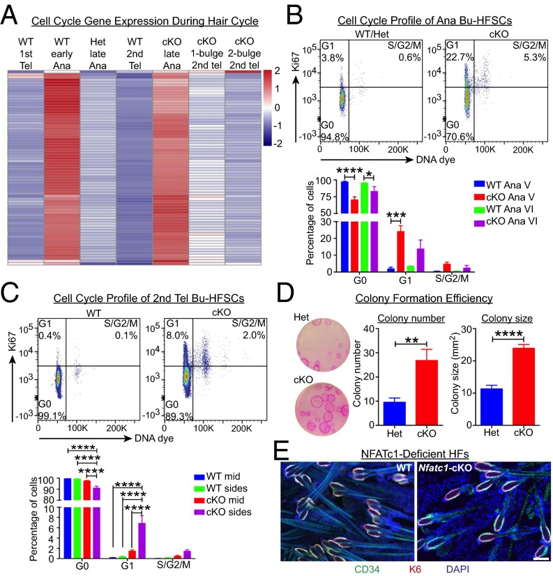
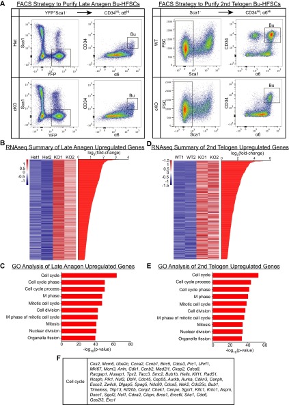
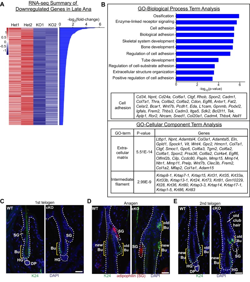
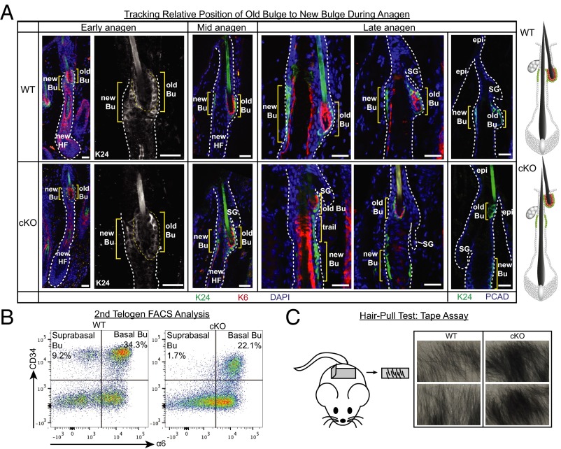
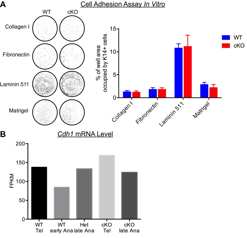
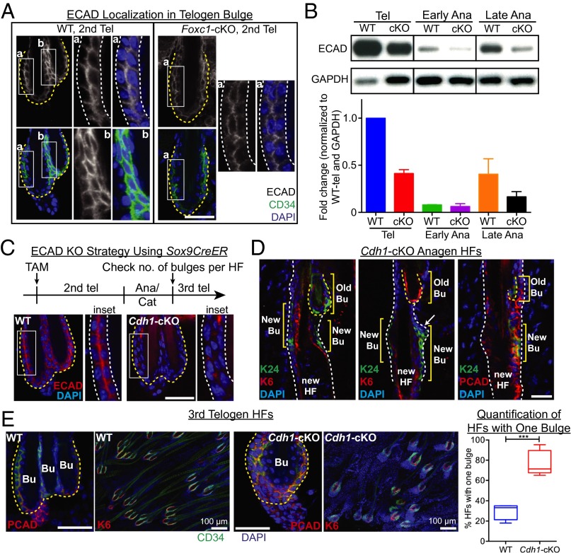
Adult tissue stem cells (SCs) reside in niches, which orchestrate SC behavior. SCs are typically used sparingly and exist in quiescence unless activated for tissue growth. Whether parsimonious SC use is essential to conserve long-term tissue-regenerating potential during normal homeostasis remains poorly understood. Here, we examine this issue by conditionally ablating a key transcription factor Forkhead box C1 (FOXC1) expressed in hair follicle SCs (HFSCs). FOXC1-deficient HFSCs spend less time in quiescence, leading to markedly shortened resting periods between hair cycles. The enhanced hair cycling accelerates HFSC expenditure, and impacts hair regeneration in aging mice. Interestingly, although FOXC1-deficient HFs can still form a new bulge that houses HFSCs for the next hair cycle, the older bulge is left unanchored. As the new hair emerges, the entire old bulge, including its reserve HFSCs and SC-inhibitory inner cell layer, is lost. We trace this mechanism first, to a marked increase in cell cycle-associated transcripts upon Foxc1 ablation, and second, to a downstream reduction in E-cadherin-mediated inter-SC adhesion. Finally, we show that when the old bulge is lost with each hair cycle, overall levels of SC-inhibitory factors are reduced, further lowering the threshold for HFSC activity. Taken together, our findings suggest that HFSCs have restricted potential in vivo, which they conserve by coupling quiescence to adhesion-mediated niche maintenance, thereby achieving long-term tissue homeostasis.

Keywords: FOXC1; aging; hair follicle; quiescence; stem cells.


Conflict of interest statement

The authors declare no conflict of interest.

Figures

Fig. S1.
Fig. S1.
Expression of FOXC1 and other key HFSC transcription factors. (A) Schematic of the adult hair cycle. HFs undergo cycles of telogen (Tel, rest), anagen (Ana, regeneration), and catagen (Cat, degeneration) throughout the lifetime of the animal. Each HF generates a new bulge and new club hair with every anagen. (B) FOXC1 expression in anagen and catagen. In anagen, FOXC1 colocalized with AE15 and GATA3, which are markers of the inner root sheath. (C) Expression of key HFSC transcription factors in WT vs. Foxc1-cKO. Note that CD34 expression in Foxc1-cKO bulge tends to be weaker than WT. (Scale bars, 30 μm.)
Fig. 1.
Fig. 1.
Depletion of FOXC1 from HFs causes faster hair cycling, yielding a sparser hair coat with age. (A) Validation of Foxc1-cKO by immunofluorescence. Abs are color-coded according to the fluorescent secondary Abs used. Bu, bulge; DP, dermal papilla; Epi, epidermis; HG, hair germ; Isth, isthmus; SG, sebaceous gland. (Scale bar, 30 μm.) (B) Foxc1-cKO mice regenerated their third hair coat much earlier than WT mice. (C) Immunofluorescence of Foxc1-cKO HF sagittal section at P50, depicting precocious Bu-HFSC activation and earlier entry into anagen than WT. (Scale bar, 30 μm.) (D) Recovered hair coats of WT (Top) and Foxc1-cKO (Bottom) were shaved repeatedly to monitor hair cycles long-term. Tel, telogen. (E) Foxc1-cKO mice underwent more frequent hair cycling and exhibited significantly shorter intervals between hair cycles. (Left) Data are mean ± SD. **P < 0.01. (Right) “Duration between hair coat recovery” refers to the time taken for > 80% of the hair coat to recover after shaving; box-and-whisker plot: midline, median; box, 25th and 75th percentiles; whiskers, minimum and maximum. ****P < 0.0001. (F) Representative example of hair coat of WT and Foxc1-cKO mice at 2 y of age. Pink box indicates zoomed-in view of lateral side of hair coat. Note visibility of skin (with some pigmented spots) underneath a sparse hair coat in Foxc1-cKO.
Fig. 2.
Fig. 2.
Without FOXC1, HFs fail to maintain multiple bulges and club hairs and display reduced HFSCs. (A) Whole-mount immunofluorescence of WT and Foxc1-cKO first telogen (Left) and second telogen (Right) HFs. CD34 (green) marks outer bulge layer (HFSCs); K6 (red) marks inner bulge layer; red autofluorescence marks club hair. Bu, bulge; HG, hair germ. [Scale bars of magnified images (Center), 30 μm.] (B) Quantification of second telogen HFs with one bulge in dorsal skin (n ≥ 4 mice, ≥80 HFs from each mouse). Box-and-whisker plot: midline, median; box, 25th and 75th percentiles; whiskers, minimum and maximum. ****P < 0.0001. (C) Quantification of total number of CD34+ HFSCs (both basal-Bu and suprabasal-Bu) per whole-mount HF (n ≥ 2 mice, ≥10 HFs per mouse). ****P < 0.0001; ns, nonsignificant. (D) Whole-mount immunofluorescence of WT and Foxc1-cKO HFs in third and fourth telogen. Note the increase in bulge numbers in WT but persistent one-bulge phenotype in Foxc1-cKO. CD34 expression is frequently weaker in Foxc1-cKO. (Scale bar, 30 μm.) (E) Whole-mount immunofluorescence of HFs in aged (≥1.5 y) WT and Foxc1-cKO animals. SG, sebaceous gland. (Scale bar, 30 μm.) (F) Sagittal section immunofluorescence of HFs which were depilated and then pulsed with BrdU for 24 h. Skin biopsies were retrieved at t = 24 and 48 h postdepilation. PCAD (P-cadherin) stains HG and outlines Bu. Note that both aged and young Foxc1-cKO HFs respond faster than WT. (Scale bar, 30 μm.) (G) Quantification of BrdU+ cells in Bu and HG 24 h postdepilation (n = 2 mice, ≥10 HFs per mouse). **P < 0.01; ****P < 0.0001. (H) Sagittal sections from day 5 postdepilated mice indicated that aged Foxc1-cKO HFs progressed to regenerate new hairs more slowly than WT. (Scale bar, 30 μm.) (I) Tracking of hair coat recovery postdepilation. Note that despite the faster response to depilation, hair coat recovery, determined by darkening of skin and appearance of new hair, was delayed in aged Foxc1-cKO mice.
Fig. S2.
Fig. S2.
FOXC1-deficient HFs can make a new bulge but fail to maintain the old one. (A) Strategy to pulse proliferating lower ORS cells with BrdU during first anagen and analyze the pattern of label-retaining cells (LRCs) in second telogen. Note that the Foxc1-cKO single bulges displayed the LRC pattern expected of a newly formed and not old bulge. Quantification shown is respective percentages of WT new bulge, WT old bulge, and Foxc1-cKO bulge that had retained BrdU label in their inner layer (n = 2 mice, 30 HFs per mouse). (B) Dyeing of first telogen hair coat and tracing it through first adult hair cycle to second telogen. Note the retention of dyed hairs in WT second telogen, but the near absence of dyed hairs in Foxc1-cKO second telogen. Immunofluorescence of second telogen HFs depicts WT old bulge anchoring old (dyed) hair, WT new bulge anchoring new (nondyed) hair, and Foxc1-cKO single bulge also anchoring a new (nondyed) hair. (C) Dyed mice were tracked closely in late anagen to monitor the fate of dyed hairs. Note that mice illustrated here entered first anagen ∼2 d later than described in Fig. S1A. (Scale bars, 30 μm.)
Fig. 3.
Fig. 3.
The old bulge contributes to HFSC quiescence. (A) Methodology of hair-shaving and hair-depilation/waxing. Note that depilation of HF removes the club hair and associated K6+ inner layer from the bulge, whereas shaving only clips away hairs at the skin surface. (B) Strategy to force WT second telogen HFs to have only one bulge. HF schematics next to mouse photos depict HF state after waxing/shaving and completion of anagen/catagen. First telogen (P19) HFs were depilated by waxing, and entered first anagen at the same time as their shaved counterparts. By second telogen (P40), shaved-HFs had two bulges/club hairs, but waxed-HFs had only one bulge/club hair. All HFs were then shaved to observe entry into second anagen. Ana, anagen; Cat, catagen; Tel, telogen. (C) Whole-mount immunofluorescence to validate strategy. Most first telogen-shaved-HFs had two bulges/club hairs, whereas first telogen-waxed HFs largely had one bulge/club hair only. (Scale bar, 100 μm.) (D, Left) qRT-PCR of SC-inhibitory factors from FACS-purified K6+ inner bulge cells. Data are mean ± SEM. (Right) Quantification of K6+ cell number per HF. Box-and-whisker plot: midline, median; box, 25th and 75th percentiles; whiskers, minimum and maximum (n ≥ 2 mice, ≥10 HFs per mouse). Note that although Bmp6 and Fgf18 were only slightly reduced on a per cell level, there were fewer K6+ inner bulge cells in FOXC1-deficient HFs, resulting in an overall reduced density of cells expressing these inhibitory factors. ****P < 0.0001. (E) Strategy to induce Foxc1-KO in second telogen two-bulge HFs using Sox9CreER. Mice were treated with tamoxifen for 5 d and observed for progression into anagen. Inset of mouse image depicts criteria to determine telogen duration, which was time taken for at least 50% of dorsal skin to enter anagen (as judged by greying, blackening or appearance of hair). (F) Second telogen duration determined by criteria described in E. WT two-bulge HFs, WT one-bulge HFs (postdepilation-recovery), Foxc1-Sox9CreER-cKO two-bulge HFs, and Foxc1-K14Cre-cKO one-bulge HFs were compared. Box-and-whisker plot: midline, median; box, 25th and 75th percentiles; whiskers, minimum and maximum (n ≥ 10 mice). **P < 0.01; ****P < 0.0001.
Fig. S3.
Fig. S3.
Elucidating the role of FOXC1 and the old bulge in hair cycling. (A) When HFs in the posterior (post) dorsal skin were forced to have one bulge, they recapitulated the precocious anagen phenotype of the anterior (ant) one-bulge HFs in Fig. 3, as evidenced by greying and darkening of posterior skin while anterior skin remained pink. (B, Left) Sox9CreER induces R26-YFP expression efficiently and specifically in HFs. (Right) qRT-PCR shows the efficient deletion of Foxc1 in FACS-purified Bu-HFSCs. Data are mean ± SEM (n = 3 mice). (C) After tamoxifen treatment in second telogen and allowing HFs to progress through anagen and third telogen, WT HFs had two or three bulges, whereas Foxc1-cKO HFs maintained their old bulges when they entered anagen precociously, but lost them by third telogen. (D) Quantification of third telogen HFs with one bulge (n ≥ 4 mice, ≥ 70 HFs from each mouse). ns, nonsignificant; ****P < 0.0001. (Scale bars, 30 μm.)
Fig. 4.
Fig. 4.
FOXC1-mediated HFSC quiescence contributes to maintaining the old bulge. (A) Heat map to illustrate changes in expression of cell cycle genes (listed in Fig. S4F) through the first hair cycle. Ana, anagen; Tel, telogen. (B) Cell cycle analysis of late anagen (substages Ana V and Ana VI) Bu-HFSCs by flow cytometry and quantification of percentages of cells in various phases of the cell cycle. Ki67 marks cycling cells; DNA content distinguishes cells in S/G2/M from G1/G0). Data are mean ± SEM (n ≥ 3 mice). *P < 0.05; ***P < 0.001; ****P < 0.0001. (C) Cell cycle analysis of second telogen Bu-HFSCs by flow cytometry and quantification of percentages of cells in various phases of the cell cycle. “Mid” and “sides” refer to midline and lateral regions of dorsal skin from which cells were analyzed. Data are mean ± SEM (n ≥ 3 mice). (D) Colony formation efficiency of Foxc1-cKO compared with Foxc1+/− Het Bu-HFSCs. (Left) Second telogen FACS-purified Bu-HFSCs were cultured in vitro for 2 wk and allowed to form colonies, which were then fixed and stained with Rhodamine B. (Center) Number of colonies formed per 33,000 cells plated. (Right) Area of each colony. Data are mean ± SEM (n ≥ 3 mice, triplicates per mouse). **P < 0.01; ****P < 0.0001. (E) Whole-mount immunofluorescence of Nfatc1-cKO HFs, showing a one-bulge phenotype. Because NFATC1 loss enhances Bu-HFSC proliferative activity, the one-bulge phenotype suggests that deregulation of quiescence may contribute to premature loss of the old bulge. (Scale bar, 100 μm.)
Fig. S4.
Fig. S4.
RNA-seq summary of up-regulated genes in Foxc1-cKO Bu-HFSCs. (A) FACS strategies to purify Bu-HFSCs from late anagen and second telogen. (B) Summary of transcriptional profiling of Foxc1-cKO vs. WT Bu-HFSCs in late anagen by RNA-seq. Shown here are significantly up-regulated genes (FPKM > 1, P < 0.05, q < 0.05). (C) Gene ontology (GO)-biological process term analysis of significantly up-regulated genes in late anagen. (D) Summary of transcriptional profiling of Foxc1-cKO vs. WT Bu-HFSCs in second telogen by RNAseq. Shown here are significantly up-regulated genes (FPKM > 1, P < 0.05, q < 0.05). (E) GO-biological process term analysis of significantly up-regulated genes in second telogen. (F) Genes in the GO-cell cycle term, commonly up-regulated in both late anagen and second telogen, are shown.
Fig. S5.
Fig. S5.
RNA-seq summary of down-regulated genes in Foxc1-cKO Bu-HFSCs, and K24 expression in HFs. (A) Summary of transcriptional profiling of Foxc1-cKO vs. WT Bu-HFSCs in late anagen by RNA-seq. Shown here are significantly down-regulated genes (FPKM > 1, P < 0.05, q < 0.05). (B) GO-biological process and cellular component term analysis of significantly down-regulated genes in late anagen. (C) K24 expression in first telogen HFs. (D) Position of old bulge, marked by K24, relative to sebaceous gland, marked by adipophilin in anagen HFs. (E) K24 expression in second telogen HFs. Note the persistence of the old bulge/club hair but its complete exclusion from the new bulge in Foxc1-cKO HF. (Scale bars, 30 μm.)
Fig. 5.
Fig. 5.
FOXC1 functions to anchor the old bulge during hair growth. (A) Immunofluorescence of sagittal sections of anagen HFs. K24 marks the old-Bu-HFSCs and the new Bu region in newly growing HFs. K6 marks the inner layer of the old Bu and the companion layer of the new HF. PCAD (P-cadherin) marks the relatively undifferentiated progenitors of the HF, including those of the bulge, outer root sheath and sebaceous gland (SG). (Scale bars, 30 μm.) (B) Flow cytometry analysis of dorsal skin epithelial cells in second telogen. Depicted are singly dissociated HF cells that were negative for SCA1 (marker of basal epidermis) and positive for CD34 (surface marker of Bu-HFSCs) and α6 integrin (surface marker of all basal epithelial cells). Note that Foxc1-cKO HFs had only CD34Hi basal Bu-HFSCs, but lacked the suprabasal-Bu-HFSC population characteristic of the interface between two bulges. (C) Tape assay. A surgical tape was affixed to the hair coat, then peeled off to assess amount of hairs that came off with the tape (n = 3 mice).
Fig. S6.
Fig. S6.
Cell–ECM adhesion assay and Cdh1 gene expression in the hair cycle. (A) In vitro cell adhesion assay. (Left) FACS-purified WT and Foxc1-cKO HFSCs were plated on collagen I, fibronectin, laminin-511, or matrigel-coated polyethylene-glycol 24-well culture plates in equal numbers in triplicates (n = 2 mice). After 1 h, nonadherent cells were washed away and adherent cells were fixed, permeabilized, and stained for keratin 14 (K14). Odyssey infrared scanner was used to visualize K14+ cells, which are depicted here as individual greyscale dots within each well (see Materials and Methods). (Right) Total area of individual adherent cells covering each well was calculated using ImageJ and presented as percentage of total well area. (B) Cdh1 transcript level from RNA-seq.
Fig. 6.
Fig. 6.
Reducing intercellular junctions between HFSCs contributes to the loss of the old bulge during new hair growth. (A) Immunofluorescence of second telogen HFs to analyze E-cadherin localization. Inset a zooms in on basal-Bu-HFSC layer in both WT and Foxc1-cKO; note that the compacted, organized bilayer of cells, characteristic of the WT bulge, is disorganized and displays extraneous cells in the Foxc1-cKO bulge. Inset b zooms in on suprabasal-Bu-HFSC layer in WT. (Scale bar, 30 μm.) (B) Immunoblotting of FACS-purified Bu-HFSCs illustrates dynamic changes in E-cadherin levels during the hair cycle. Quantifications are mean ± SEM of ≥ 3 independent replicates normalized to WT-tel using GAPDH as loading control. Note that FOXC1 loss reduces overall E-cadherin levels but does not alter their dynamics during the hair cycle. (C) Strategy to ablate Cdh1 gene expression in HFs by using Sox9-CreER mice. Immunofluorescence images depict loss of E-cadherin after tamoxifen treatment and appearance of disorganized cells within the bulge, compared with WT. (Scale bar, 30 μm.) (D) Immunofluorescence of sagittal sections of Cdh1-cKO anagen HFs depicting the position of the old bulge relative to the newly specified bulge. Arrow points to K24+ cells left behind as the old bulge moved upwards. (Scale bar, 30 μm.) (E) Immunofluorescence of WT and Cdh1-cKO third telogen HFs. Quantifications show that most Cdh1-cKO HFs had only one bulge by their third telogen. Box-and-whisker plot: midline, median; box, 25th and 75th percentiles; whiskers, minimum and maximum (n ≥ 4 mice, ≥80 HFs per mouse). (Scale bars, 30 μm unless indicated otherwise.) ***P < 0.001.

References

    1. Orford KW, Scadden DT. Deconstructing stem cell self-renewal: Genetic insights into cell-cycle regulation. Nat Rev Genet. 2008;9(2):115–128. - PubMed
    1. Cheung TH, Rando TA. Molecular regulation of stem cell quiescence. Nat Rev Mol Cell Biol. 2013;14(6):329–340. - PMC - PubMed
    1. Morrison SJ, Spradling AC. Stem cells and niches: Mechanisms that promote stem cell maintenance throughout life. Cell. 2008;132(4):598–611. - PMC - PubMed
    1. Hsu YC, Fuchs E. A family business: Stem cell progeny join the niche to regulate homeostasis. Nat Rev Mol Cell Biol. 2012;13(2):103–114. - PMC - PubMed
    1. Chen CC, et al. Organ-level quorum sensing directs regeneration in hair stem cell populations. Cell. 2015;161(2):277–290. - PMC - PubMed

Publication types