. 2016 Jun 1;115(5):2434-45.
doi: 10.1152/jn.00737.2015. Epub 2016 Feb 24.

Role of Ih in differentiating the dynamics of the gastric and pyloric neurons in the stomatogastric ganglion of the lobster, Homarus americanus

Affiliations

Role of Ih in differentiating the dynamics of the gastric and pyloric neurons in the stomatogastric ganglion of the lobster, Homarus americanus

Lin Zhu et al. J Neurophysiol. .

Abstract

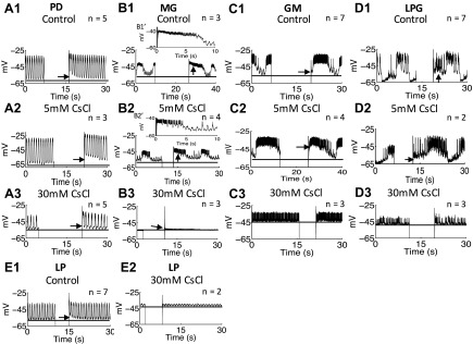
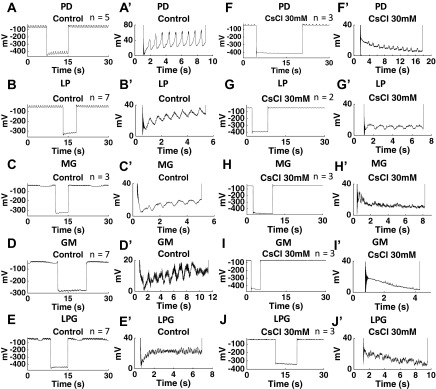
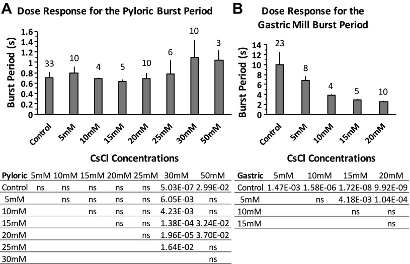
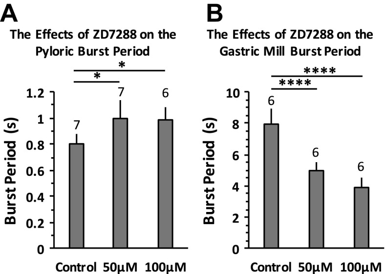
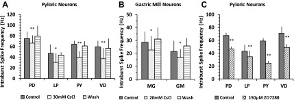
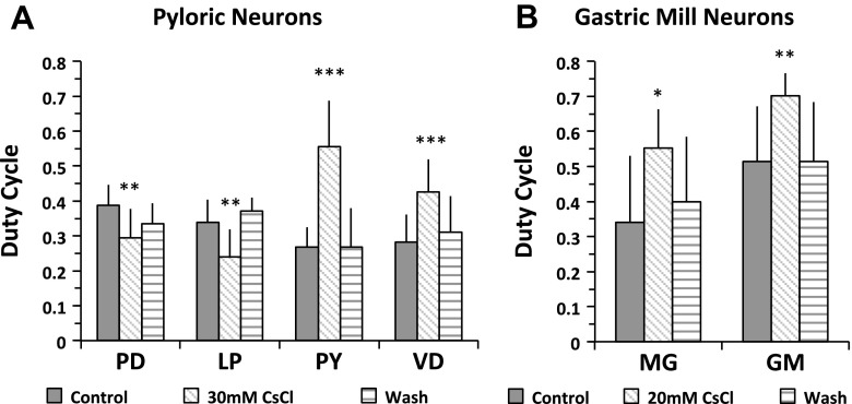
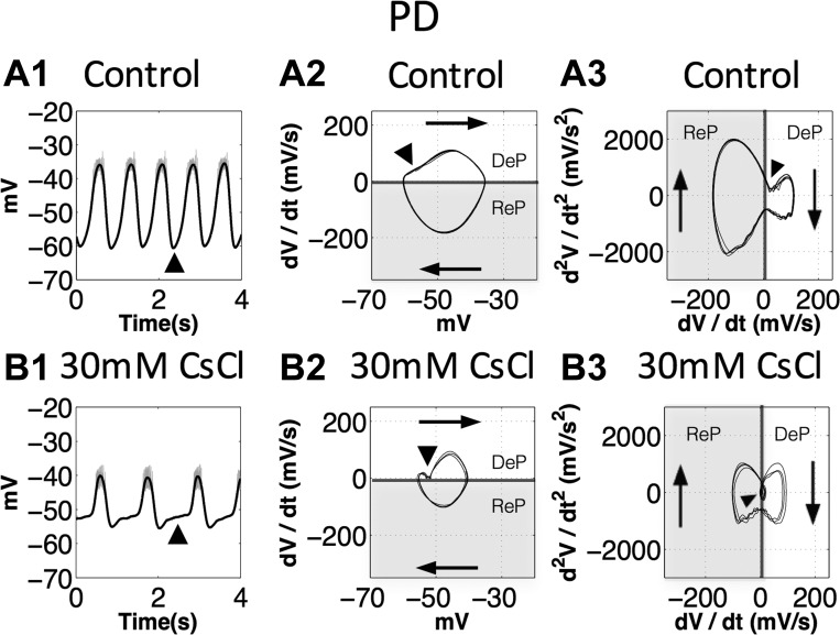
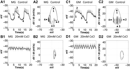
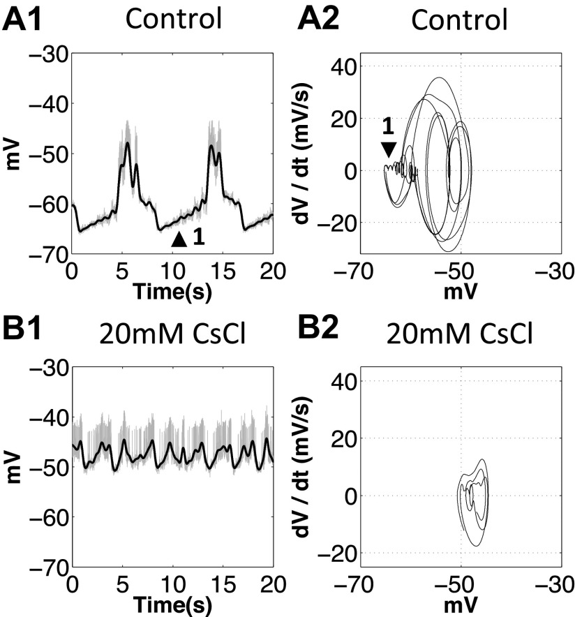
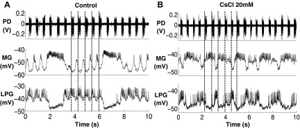
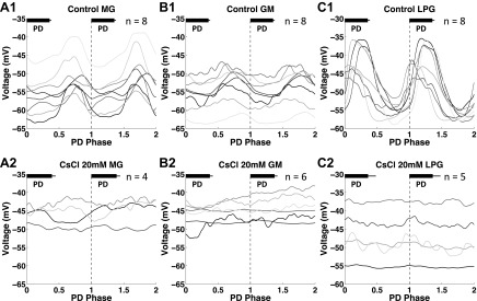
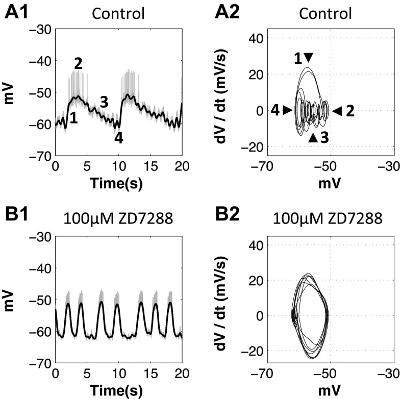
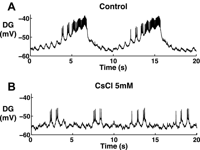
The hyperpolarization-activated inward cationic current (Ih) is known to regulate the rhythmicity, excitability, and synaptic transmission in heart cells and many types of neurons across a variety of species, including some pyloric and gastric mill neurons in the stomatogastric ganglion (STG) in Cancer borealis and Panulirus interruptus However, little is known about the role of Ih in regulating the gastric mill dynamics and its contribution to the dynamical bifurcation of the gastric mill and pyloric networks. We investigated the role of Ih in the rhythmic activity and cellular excitability of both the gastric mill neurons (medial gastric, gastric mill) and pyloric neurons (pyloric dilator, lateral pyloric) in Homarus americanus Through testing the burst period between 5 and 50 mM CsCl, and elimination of postinhibitory rebound and voltage sag, we found that 30 mM CsCl can sufficiently block Ih in both the pyloric and gastric mill neurons. Our results show that Ih maintains the excitability of both the pyloric and gastric mill neurons. However, Ih regulates slow oscillations of the pyloric and gastric mill neurons differently. Specifically, blocking Ih diminishes the difference between the pyloric and gastric mill burst periods by increasing the pyloric burst period and decreasing the gastric mill burst period. Moreover, the phase-plane analysis shows that blocking Ih causes the trajectory of slow oscillations of the gastric mill neurons to change toward the pyloric sinusoidal-like trajectories. In addition to regulating the pyloric rhythm, we found that Ih is also essential for the gastric mill rhythms and differentially regulates these two dynamics.

Keywords: central pattern generator; hyperpolarization-activated inward cationic current.


Figures

Fig. 1.
Fig. 1.
Postinhibitory rebound (PIR) in the pyloric dilator (PD), medial gastric (MG), lateral pyloric (LP), gastric mill (GM), and lateral posterior gastric (LPG) neurons was blocked by 30 mM CsCl. Arrowheads show PIR after releasing from injecting 10-nA hyperpolarizing current. A1: the PIR was present in the PD neurons in the control condition. A2: the PIR was present in the PD neurons after application of 5 mM CsCl. A3: the PIR was blocked in the PD neurons after application of 30 mM CsCl. B1′ and B2′ are magnified images to show PIR in B1 and B2, respectively. B1 and B1′: the PIR was present in the MG neuron in the control condition and caused the temporary increase in burst duration and inraburst spike frequency. B2 and B2′: the PIR was present in the MG neuron after application of 5 mM CsCl and caused the temporary increase in burst duration and intraburst spike frequency. B3: the PIR was blocked in the MG neuron after application of 30 mM CsCl. C1: PIR was present in the control condition in the GM neurons. C2: PIR was present in the GM neurons after application of 5 mM CsCl. C3: the PIR was blocked in the GM neurons after application of 30 mM CsCl. D1: the PIR was present in the LPG neurons in the control condition. D2: the PIR was present in the LPG neurons after application of 5 mM CsCl. D3: the PIR was blocked in the LPG neurons after application of 30 mM CsCl. E1: the PIR was present in the LP neuron in the control condition. E2: PIR was blocked in the LP neuron after application of 30 mM CsCl. The number of trials (n) is indicated.
Fig. 2.
Fig. 2.
CsCl (30 mM) eliminated the voltage “sag” induced by hyperpolarization-activated inward cationic current (Ih) in the PD, LP, MG, GM, and LP neurons. A′, B′, C′, D′, E′, F′, G′, H′, I′, and J′ are magnified images from A, B, C, D, E, F, G, H, I, and J, respectively. A, A′, B, B′, C, C′, D, D′, E, E′: in the control condition, the PD, LP, MG, GM and LPG neurons all showed voltage sag after a −10-nA hyperpolarization. F, F′, G, G′, H, H′, I, I′, J, J′: after application of 30 mM CsCl, the PD, LP, MG, GM, and LPG neurons did not show voltage sag after a −10-nA hyperpolarization. n Values are indicated in each plot. The large negative voltage value during hyperpolarization (A-J) was due to the unbalanced bridge when using the single microelectrode recording. Balancing the bridge is an adjustment of the output signal; it has no effect on the cells.
Fig. 3.
Fig. 3.
Changes of the burst period of pyloric and gastric neurons after blocking Ih with a series of concentrations of CsCl ranging from 5 to 50 mM. A: the increase in the pyloric burst period reached significance at application of 30 and 50 mM CsCl. One-way ANOVA followed by Tukey's honest significant difference (HSD) post hoc test, F(7,73) = 7.981, P < 0.0001. B: the gastric mill burst period decreased after application of CsCl at 5, 10, 15, and 20 mM concentrations. One-way ANOVA followed by Tukey's HSD post hoc test, F(4,45) = 38.654, P < 0.0001. ns, Nonsignificant. Error bars are represented as SD. The no. of trials (n) is indicated.
Fig. 4.
Fig. 4.
Blocking Ih with ZD-7288 increased the pyloric burst period and decreased the gastric mill burst period. A: the pyloric burst period increased after 50 and 100 μM ZD-7288 applications. One-way ANOVA followed by Tukey's HSD post hoc test, F(2,17) = 6.933, P = 0.006. B: the gastric mill burst period decreased after 50 and 100 μM ZD-7288 applications. One-way ANOVA followed by Tukey's HSD post hoc test, F(2,15) = 49.546, P < 0.0001. Error bars are represented as SD. The no. of trials (n) is indicated. *P < 0.05 and ****P < 0.00001.
Fig. 5.
Fig. 5.
Blocking Ih decreased the intraburst spike frequency of both pyloric and gastric neurons. A: the intraburst spike frequency of the pyloric neurons decreased after application of 30 mM CsCl, paired Student's t-test, PD: n = 10, P = 0.006; LP: n = 7, P = 0.033; ventricular dilator (VD): n = 7, P = 0.003; pyloric (PY): n = 7, P = 0.007. B: the intraburst spike frequency of the gastric mill neurons decreased after application of 20 mM CsCl, paired Student's t-test, MG: n = 9, P = 0.024; GM: n = 10, P = 0.019. C: the intraburst spike frequency of the pyloric neurons decreased after application of 150 μM ZD-7288, paired Student's t-test, PD: n = 3, P = 0.005; PY: n = 3, P = 0.001; LP: n = 3, P = 0.001; VD: n = 3, P = 0.024. A, B, and C: *P < 0.05 and **P < 0.01. Error bars are represented SD.
Fig. 6.
Fig. 6.
Blocking Ih altered the duty cycle of the pyloric and gastric mill neurons. A: the duty cycle of the PD and LP neurons decreased, and the duty cycle of the PY and VD neurons increased, paired Student's t-test, PD: n = 10, P = 0.003; LP: n = 7, P = 0.004; PY: n = 7, P = 0.0003; VD: n = 7, P = 0.0003. B: the duty cycle of the MG and GM neurons increased, paired Student's t-test, MG: n = 9, P = 0.015; GM: n = 10, P = 0.001. A and B: *P < 0.05, **P < 0.01, and ***P < 0.001. Error bars are represented as SD.
Fig. 7.
Fig. 7.
The second derivative of voltage (d2V/dt2) at the onset of the depolarizing phase of the PD neurons decreased when Ih was blocked. DeP, depolarization phase; ReP, repolarization phase. The shaded sections show the repolarization phases. Arrowheads indicate the onset of the depolarizing phase. A1 and B1: black traces, the slow oscillations; gray traces, the original voltage traces. A2 and B2: voltage vs. the first derivative of voltage (dV/dt) diagrams, traces travel clockwise, dV/dt at the onset of the depolarizing phase decreased after application of 30 mM CsCl. A3 and B3: dV/dt vs. d2V/dt2 diagrams, black arrowhead shows d2V/dt2 at the onset of the depolarizing phase. Note that, after the onset, application of 30 mM CsCl (B3) causes the rate of depolarization to decelerate (d2V/dt2 < 0), leading to a much slower depolarization phase (B2 and B1).
Fig. 8.
Fig. 8.
When Ih was blocked with 20 mM CsCl, the oscillatory trajectory of the MG and GM neurons became pyloric like. A1, A2, C1, and C2: 1, depolarizing phase; 2, plateau phase; 3, repolarizing phase; 4, valley. A2, B2, C2, and D2: after application of 20 mM CsCl, the oscillatory trajectory of the MG and GM neurons changed toward sinusoidal like, and the pyloric modulation was diminished. A1, B1, C1, and D1: gray traces show the orignal voltage traces, and black traces show the slow oscillations after filtering.
Fig. 9.
Fig. 9.
When Ih was blocked with 20 mM CsCl, the oscillatory trajectory of the LPG neuron changed. A1 and A2: 1, voltage valleys. After application of 20 mM CsCl, the pyloric modulation was diminished.
Fig. 10.
Fig. 10.
Pyloric modulation of the MG and LPG neurons diminished after application of 20 mM CsCl. The dark gray lines indicate the onset of PD bursts. A: the PD modulation was present in the MG and LPG oscillations in the control condition. B: the PD modulation was no longer present in the MG and LPG oscillations after application of 20 mM CsCl, n = 4.
Fig. 11.
Fig. 11.
PD modulation on the MG, GM, and LPG neurons diminished after Ih was blocked with 20 mM CsCl. Black bars in each graph represent the PD burst in each burst cycle. A1: in the control condition, the MG interburst membrane potential was hyperpolarized during PD bursts and depolarized during PD interburst intervals. A2: after application of 20 mM CsCl, the MG interburst membrane potential no longer oscillated with the PD phase. B1: in the control condition, the GM interburst membrane potential was hyperpolarized during PD bursts and depolarized during PD interburst intervals. B2: after application of 20 mM CsCl, the GM interburst membrane potential no longer oscillated with the PD phase. C1: in the control condition, the LPG burst membrane potential was depolarized during PD bursts and hyperpolarized during PD interburst intervals. B2: after application of 20 mM CsCl, the LPG burst membrane potential no longer oscillated with the PD phase.
Fig. 12.
Fig. 12.
When Ih was blocked with 100 μM ZD-7288, the oscillatory trajectory of the GM neurons became pyloriclike. A1 and A2: 1, depolarizing phase; 2, plateau phase; 3, repolarizing phase; 4, valley. B1 and B2: after application of 100 μM ZD-7288, the oscillatory trajectory neurons changed toward sinusoidal like, and the pyloric modulation was diminished.
Fig. 13.
Fig. 13.
When blocking Ih with 5 mM CsCl, the burst period, burst duration, and intraburst spike frequency of the dorsal gastric (DG) neuron decreased, n = 2.

Similar articles

Cited by

References

    1. Aponte Y, Lien CC, Reisinger E, Jonas P. Hyperpolarization-activated cation channels in fast-spiking interneurons of rat hippocampus. J Physiol 574: 229–243, 2006. - PMC - PubMed
    1. Ballo AW, Bucher D. Complex intrinsic membrane properties and dopamine shape spiking activity in a motor axon. J Neurosci 29: 5062–5074, 2009. - PMC - PubMed
    1. Bartos M, Manor Y, Nadim F, Marder E, Nusbaum MP. Coordination of fast and slow rhythmic neuronal circuits. J Neurosci 19: 6650–6660, 1999. - PMC - PubMed
    1. Beaumont V, Zucker RS. Enhancement of synaptic transmission by cyclic AMP modulation of presynaptic Ih channels. Nat Neurosci 3: 133–141, 2000. - PubMed
    1. Brown DA, Adams PR. Muscarinic suppression of a novel voltage-sensitive K+ current in a vertebrate neurone. Nature 283: 673–676, 1980. - PubMed

Publication types

Substances

LinkOut - more resources