

Longitudinal evidence for anterograde trans-synaptic degeneration after optic neuritis
- PMID: 26912640
- PMCID: PMC5839599
- DOI: 10.1093/brain/awv396
Longitudinal evidence for anterograde trans-synaptic degeneration after optic neuritis
Abstract
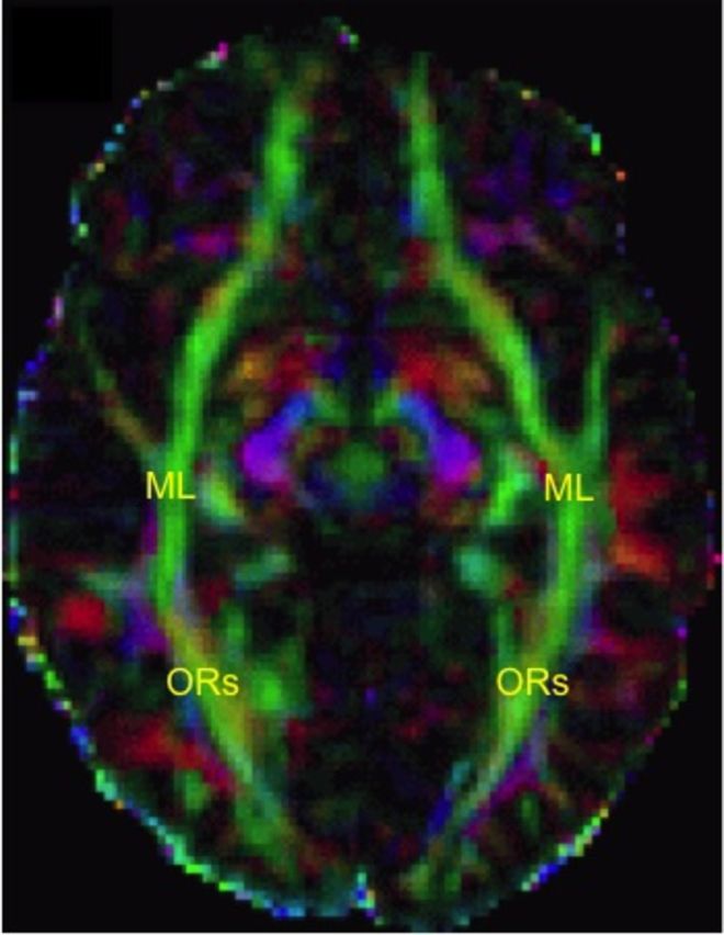
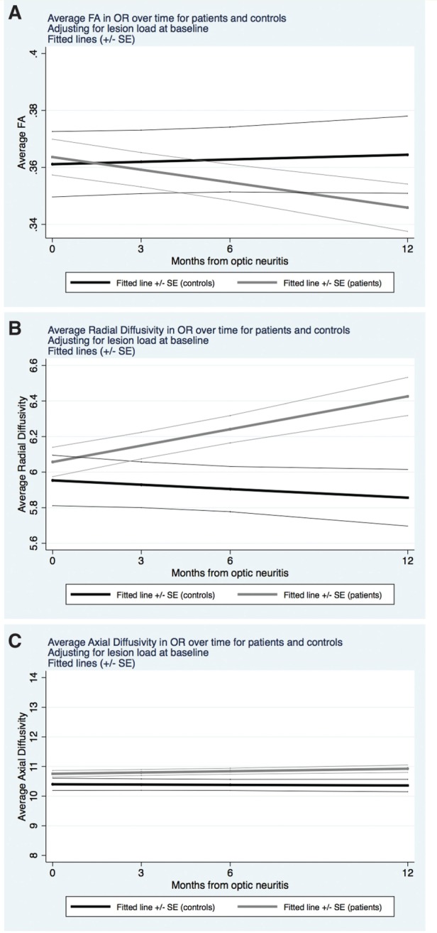
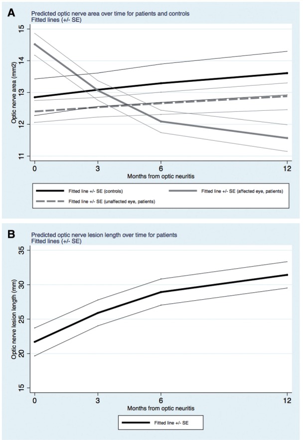
In multiple sclerosis, microstructural damage of normal-appearing brain tissue is an important feature of its pathology. Understanding these mechanisms is vital to help develop neuroprotective strategies. The visual pathway is a key model to study mechanisms of damage and recovery in demyelination. Anterograde trans-synaptic degeneration across the lateral geniculate nuclei has been suggested as a mechanism of tissue damage to explain optic radiation abnormalities seen in association with demyelinating disease and optic neuritis, although evidence for this has relied solely on cross-sectional studies. We therefore aimed to assess: (i) longitudinal changes in the diffusion properties of optic radiations after optic neuritis suggesting trans-synaptic degeneration; (ii) the predictive value of early optic nerve magnetic resonance imaging measures for late optic radiations changes; and (iii) the impact on visual outcome of both optic nerve and brain post-optic neuritis changes. Twenty-eight consecutive patients with acute optic neuritis and eight healthy controls were assessed visually (logMAR, colour vision, and Sloan 1.25%, 5%, 25%) and by magnetic resonance imaging, at baseline, 3, 6, and 12 months. Magnetic resonance imaging sequences performed (and metrics obtained) were: (i) optic nerve fluid-attenuated inversion-recovery (optic nerve cross-sectional area); (ii) optic nerve proton density fast spin-echo (optic nerve proton density-lesion length); (iii) optic nerve post-gadolinium T1-weighted (Gd-enhanced lesion length); and (iv) brain diffusion-weighted imaging (to derive optic radiation fractional anisotropy, radial diffusivity, and axial diffusivity). Mixed-effects and multivariate regression models were performed, adjusting for age, gender, and optic radiation lesion load. These identified changes over time and associations between early optic nerve measures and 1-year global optic radiation/clinical measures. The fractional anisotropy in patients' optic radiations decreased (P = 0.018) and radial diffusivity increased (P = 0.002) over 1 year following optic neuritis, whereas optic radiation measures were unchanged in controls. Also, smaller cross-sectional areas of affected optic nerves at 3 months post-optic neuritis predicted lower fractional anisotropy and higher radial diffusivity at 1 year (P = 0.007) in the optic radiations, whereas none of the inflammatory measures of the optic nerve predicted changes in optic radiations. Finally, greater Gd-enhanced lesion length at baseline and greater optic nerve proton density-lesion length at 1 year were associated with worse visual function at 1 year (P = 0.034 for both). Neither the cross-sectional area of the affected optic nerve after optic neuritis nor the damage in optic radiations was associated with 1-year visual outcome. Our longitudinal study shows that, after optic neuritis, there is progressive damage to the optic radiations, greater in patients with early residual optic nerve atrophy, even after adjusting for optic radiation lesions. These findings provide evidence for trans-synaptic degeneration.
Keywords: diffusion tensor imaging; multiple sclerosis; optic neuritis; optic radiations; trans-synaptic degeneration.
© The Author (2016). Published by Oxford University Press on behalf of the Guarantors of Brain. All rights reserved. For Permissions, please email: journals.permissions@oup.com.
Figures
References
-
- Audoin B, Fernando KT, Swanton JK, Thompson AJ, Plant GT, Miller DH . Selective magnetization transfer ratio decrease in the visual cortex following optic neuritis . Brain 2006. ; 129 ( Pt 4 ): 1031 – 9 . - PubMed
-
- Balk LJ, Steenwijk MD, Tewarie P, Daams M, Killestein J, Wattjes MP, et al. . Bidirectional trans-synaptic axonal degeneration in the visual pathway in multiple sclerosis . J Neurol Neurosurg Psychiatry 2015. ; 86 : 419 – 24 . - PubMed
-
- Basser PJ, Mattiello J, LeBihan D . Estimation of the effective self-diffusion tensor from the NMR spin echo . J Magn Reson B 1994. ; 103 : 247 – 54 . - PubMed
-
- Beaulieu C . The basis of anisotropic water diffusion in the nervous system: a technical review . NMR Biomed 2002. ; 15 : 435 – 55 . - PubMed
Publication types
MeSH terms
LinkOut - more resources
Full Text Sources
Other Literature Sources
