

Network-selective vulnerability of the human cerebellum to Alzheimer's disease and frontotemporal dementia
- PMID: 26912642
- PMCID: PMC5839595
- DOI: 10.1093/brain/aww003
Network-selective vulnerability of the human cerebellum to Alzheimer's disease and frontotemporal dementia
Erratum in
-
Corrigendum.Brain. 2016 Nov 1;139(11):e66. doi: 10.1093/brain/aww191. Brain. 2016. PMID: 29106483 Free PMC article. No abstract available.
Abstract
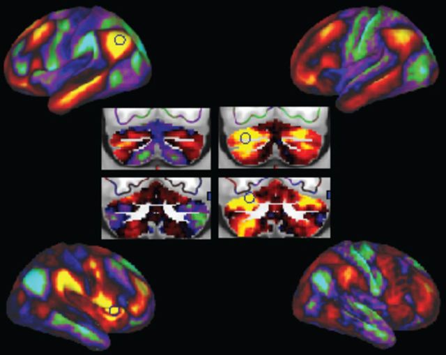
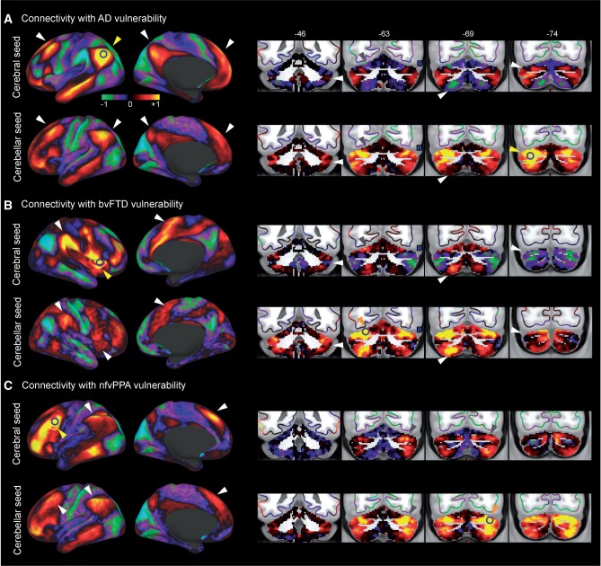
SEE SCHMAHMANN DOI101093/BRAIN/AWW064 FOR A SCIENTIFIC COMMENTARY ON THIS ARTICLE: Neurodegenerative diseases are associated with distinct and distributed patterns of atrophy in the cerebral cortex. Emerging evidence suggests that these atrophy patterns resemble intrinsic connectivity networks in the healthy brain, supporting the network-based degeneration framework where neuropathology spreads across connectivity networks. An intriguing yet untested possibility is that the cerebellar circuits, which share extensive connections with the cerebral cortex, could be selectively targeted by major neurodegenerative diseases. Here we examined the structural atrophy in the cerebellum across common types of neurodegenerative diseases, and characterized the functional connectivity patterns of these cerebellar atrophy regions. Our results showed that Alzheimer's disease and frontotemporal dementia are associated with distinct and circumscribed atrophy in the cerebellum. These cerebellar atrophied regions share robust and selective intrinsic connectivity with the atrophied regions in the cerebral cortex. These findings for the first time demonstrated the selective vulnerability of the cerebellum to common neurodegenerative disease, extending the network-based degeneration framework to the cerebellum. Our work also has direct implications on the cerebellar contribution to the cognitive and affective processes that are compromised in neurodegeneration as well as the practice of using the cerebellum as reference region for ligand neuroimaging studies.
Keywords: cerebellum; intrinsic connectivity; neurodegeneration; selective vulnerability.
© The Author (2016). Published by Oxford University Press on behalf of the Guarantors of Brain. All rights reserved. For Permissions, please email: journals.permissions@oup.com.
Figures


Comment in
-
Dementia: Cerebellar atrophy has disease-specific patterns.Nat Rev Neurol. 2016 Apr;12(4):188. doi: 10.1038/nrneurol.2016.28. Epub 2016 Mar 11. Nat Rev Neurol. 2016. PMID: 26965672 No abstract available.
-
Cerebellum in Alzheimer's disease and frontotemporal dementia: not a silent bystander.Brain. 2016 May;139(Pt 5):1314-8. doi: 10.1093/brain/aww064. Brain. 2016. PMID: 27189578 No abstract available.
-
C9orf72 mutations and the puzzle of cerebro-cerebellar network degeneration.Brain. 2016 Aug;139(Pt 8):e44. doi: 10.1093/brain/aww103. Epub 2016 May 3. Brain. 2016. PMID: 27190011 Free PMC article. No abstract available.
-
Reply: C9orf72 mutations and the puzzle of cerebro-cerebellar network degeneration.Brain. 2016 Aug;139(Pt 8):e45. doi: 10.1093/brain/aww104. Epub 2016 May 3. Brain. 2016. PMID: 27190029 No abstract available.
-
The Crus exhibits stronger functional connectivity with executive network nodes than with the default mode network.Brain. 2018 Apr 1;141(4):e24. doi: 10.1093/brain/awy025. Brain. 2018. PMID: 29471474 No abstract available.
-
Reply: The Crus exhibits stronger functional connectivity with executive network nodes than with the default mode network.Brain. 2018 Apr 1;141(4):e25. doi: 10.1093/brain/awy041. Brain. 2018. PMID: 29471509 No abstract available.
References
Publication types
MeSH terms
Grants and funding
LinkOut - more resources
Full Text Sources
Other Literature Sources
Medical
