. 2016 Mar;139(Pt 3):807-15.
doi: 10.1093/brain/awv398. Epub 2016 Feb 8.

The topograpy of demyelination and neurodegeneration in the multiple sclerosis brain

Affiliations

The topograpy of demyelination and neurodegeneration in the multiple sclerosis brain

Lukas Haider et al. Brain. 2016 Mar.

Abstract

Multiple sclerosis is a chronic inflammatory disease with primary demyelination and neurodegeneration in the central nervous system. In our study we analysed demyelination and neurodegeneration in a large series of multiple sclerosis brains and provide a map that displays the frequency of different brain areas to be affected by these processes. Demyelination in the cerebral cortex was related to inflammatory infiltrates in the meninges, which was pronounced in invaginations of the brain surface (sulci) and possibly promoted by low flow of the cerebrospinal fluid in these areas. Focal demyelinated lesions in the white matter occurred at sites with high venous density and additionally accumulated in watershed areas of low arterial blood supply. Two different patterns of neurodegeneration in the cortex were identified: oxidative injury of cortical neurons and retrograde neurodegeneration due to axonal injury in the white matter. While oxidative injury was related to the inflammatory process in the meninges and pronounced in actively demyelinating cortical lesions, retrograde degeneration was mainly related to demyelinated lesions and axonal loss in the white matter. Our data show that accumulation of lesions and neurodegeneration in the multiple sclerosis brain does not affect all brain regions equally and provides the pathological basis for the selection of brain areas for monitoring regional injury and atrophy development in future magnetic resonance imaging studies.

Keywords: cerebral arteries; cerebral veins; demyelination; multiple sclerosis; neurodegeneration.


Figures

None
Multiple sclerosis is characterized by widespread primary demyelination and progressive degeneration, driven by heterogeneous mechanisms. Haider et al. provide a topographic map of the frequency with which different brain regions are affected by these processes, and show that demyelination and neurodegeneration involve inflammatory as well as vascular changes.
Figure 1
Figure 1
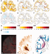
Probability maps of multiple sclerosis patients. Probability maps of multiple sclerosis patients ( A , C and E ) and age-matched controls ( B , D and F ). The colour codes indicate the probability of finding lesions (in % of cases A and B ), the cumulative frequencies of neurons affected by retrograde neurodegeneration ( C / D ) and the cumulative frequencies of meningeal inflammatory cells ( E / F ) in a specific location of this virtual brain slice. ( A ) The probability of demyelinating lesions in the white and grey matter of multiple sclerosis patients. ( B ) The probability of leukoaraiosis in control subjects. ( C ) The cumulative number of neurons with retrograde degeneration in multiple sclerosis patients. ( E ) The accumulated number of inflammatory cells in the leptomeninges of multiple sclerosis patients. The highest incidence of demyelination ( A ) in the white matter is seen in the so-called watershed areas, which are located at the borders of the supply territories of the major cerebral arteries. In contrast, cortical lesions are mainly concentrated in invaginations of the cortical surface, such as the cortical sulci, in regions with high incidence of meningeal inflammatory infiltrates ( E ). Retrograde neurodegeneration is mainly seen in the deep cortical layers and the deep grey matter and in part follows the putative fibre projection from white matter lesions into the cortex ( C ). In age-matched controls no plaques of primary demyelination were present, but there were areas of diffuse white matter alterations (leukoaraiosis) in 43% of the cases investigated (blue areas in B ). Cortical neurodegeneration was much less pronounced compared to that seen in multiple sclerosis ( D ), but showed a topographically similar distribution compared to that seen in multiple sclerosis patients. Inflammatory infiltrates were also seen in low numbers and incidence in the meninges of the age-matched controls ( F ). Definitions of regions of interest: ( G ) venous density atlas from Grabner et al. (2014) depicts the density of veins (red) in different brain areas. Brains were scanned in a 7 T MRI and a venous map of the normal human brain was created. ( H ) Turnbull staining showing the iron distribution throughout the brain. ( I ) Our regions of interest in one hemisphere of the virtual brain map. Areas of low CSF flow are indicated by blue, watershed area in purple and the basal ganglia in pink.
Figure 2
Figure 2
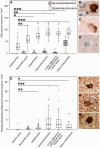
Neurodegeneration in the multiple sclerosis cortex. Oxidative injury in cortical neurons is reflected by two different neuronal changes. ( A–D ) The first is the intense and diffuse immunoreactivity for oxidized phospholipids (E06 immunoreactivity) in neurons ( B ), associated with beading and fragmentation of cell processes (indicated by the arrowhead) and neuronal apoptosis ( Fischer et al. , 2013 ). This is mainly seen in active cortical lesions, but in lower frequency also in inactive lesions and the normal-appearing multiple sclerosis cortex (grey bars in A ). The second is reflected by a granular, lipofuscin-like immunoreactivity in neurons ( C ). This is significantly more frequent (white bars in A ) in the multiple sclerosis cortex compared to controls, but within multiple sclerosis its incidence is not significantly different between active and inactive lesions. ( D ) A normal neuron without immunoreactivity for oxidized phospholipids in the multiple sclerosis cortex. ( E–H ) Retrograde neurodegeneration is reflected by the cytoplasmic accumulation of phosphorylated neurofilaments ( F ). This is frequently associated with ballooning of neurons and an eccentric location of the nerve cell nucleus ( G and H ). Retrograde neurodegeneration is most prominently seen in the cortex adjacent to demyelinated lesions with axonal transection in the white matter ( E ). * P < 0.05; ** P < 0.01; *** P <0.001.

References

    1. Abbott NJ . Evidence for bulk flow of brain interstitial fluid: significance for physiology and pathology . Neurochem Int 2004. ; 45 : 545 – 52 . - PubMed
    1. Aboul-Enein F, Rauschka H, Kornek B, Stadelmann C, Stefferl A, Bruck W, et al. . Preferential loss of myelin-associated glycoprotein reflects hypoxia-like white matter damage in stroke and inflammatory brain diseases . J Neuropathol Exp Neurol 2003. ; 62 : 25 – 33 . - PubMed
    1. Bo L, Vedeler CA, Nyland H, Trapp BD, Mork SJ . Intracortical multiple sclerosis lesions are not associated with increased lymphocyte infiltration . Mult Scler 2003. ; 9 : 323 – 31 . - PubMed
    1. Brown WR, Thore CR . Review: cerebral microvascular pathology in ageing and neurodegeneration . Neuropathol Appl Neurobiol 2011. ; 37 : 56 – 74 . - PMC - PubMed
    1. Brownell B, Hughes JT . The distribution of plaques in the cerebrum in multiple sclerosis . J Neurol Neurosurg Psychiatry 1962. ; 25 : 315 – 20 . - PMC - PubMed

Publication types