. 2016 Feb 5;351(6273):582-6.
doi: 10.1126/science.aad2080.

Diamond family of nanoparticle superlattices

Affiliations

Diamond family of nanoparticle superlattices

Wenyan Liu et al. Science. .

Abstract

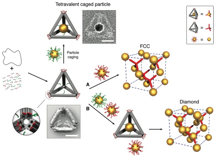
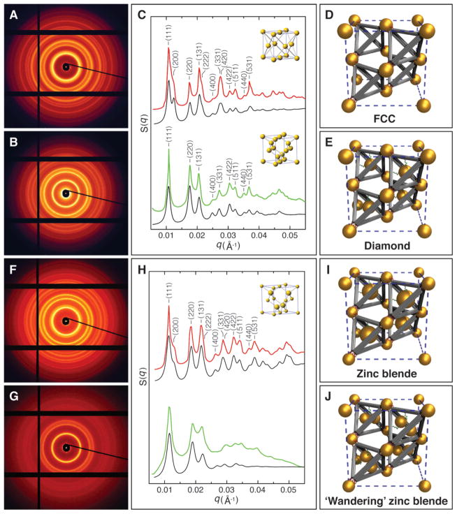
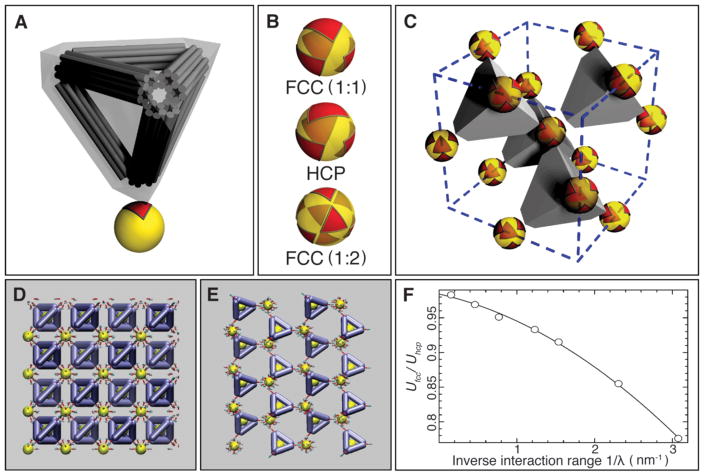
Diamond lattices formed by atomic or colloidal elements exhibit remarkable functional properties. However, building such structures via self-assembly has proven to be challenging because of the low packing fraction, sensitivity to bond orientation, and local heterogeneity. We report a strategy for creating a diamond superlattice of nano-objects via self-assembly and demonstrate its experimental realization by assembling two variant diamond lattices, one with and one without atomic analogs. Our approach relies on the association between anisotropic particles with well-defined tetravalent binding topology and isotropic particles. The constrained packing of triangular binding footprints of truncated tetrahedra on a sphere defines a unique three-dimensional lattice. Hence, the diamond self-assembly problem is solved via its mapping onto two-dimensional triangular packing on the surface of isotropic spherical particles.


Figures

Fig. 1
Fig. 1. Schematic illustration of the experimental strategy
A circular single-stranded M13 DNA genome is folded by a set of helper strands to generate a rigid tetrahedral DNA origami cage containing two sets of sticky-ended DNA strands. One set (green) is projected from the inner faces of the edges, functioning as an anchor to encapsulate and hold the guest particle (uniformly coated with green strands) inside the cage; another set (red) is installed at each vertex of the tetrahedral cage, acting as a sticky patch to provide binding to the basis particles (uniformly coated with red strands). Zoom in: a detailed view of the vertex (truncated) of the tetrahedron cage; image blow the tetrahedron model: a reconstructed cryo-EM density map of the tetrahedron. The guest and the basis particles coated with corresponding complementary DNA can either individually interact with the tetrahedral cages to form tetravalent caged particles and FCC superlattices (route A, empty cages) respectively, or together hybridize with the tetrahedral cages to create diamond crystals (route B, with caged particle). A representative of a constructed tetravalent caged particle is shown in a negative-staining TEM image beside the model. Top right box is a visual definition of the system components for simplified illustration of shown FCC and diamond superlattices. Scale bars, 20 nm.
Fig. 2
Fig. 2. SAXS characterization of the diamond family of nanoparticle superlattices
(A) 2D SAXS pattern of the FCC superlattices constructed with basis particles (core diameter 14.5 nm) and tetrahedral DNA origami cages. (B) 2D SAXS pattern of the diamond superlattices formed from basis particles (core diameter 14.5 nm) and tetravalent caged particles (core diameter 14.5 nm). (C) Integrated 1D patterns. Top channel: Experimental (red) and calculated (black) 1D SAXS patterns for the FCC crystals. Bottom channel: Experimental (green) and calculated (black) 1D SAXS patterns for the diamond crystals. Insets are standard FCC and diamond unit cells respectively. (D) Unit cell model of the assembled FCC superlattice. (E) Unit cell model of the constructed diamond crystal. (F) 2D SAXS pattern of the zinc blende lattices constructed with basis particles (core diameter 8.7 nm) and tetravalent caged particles (core diameter 14.5 nm). (G) 2D SAXS pattern of the ‘wandering’ zinc blende lattices formed from basis particles (core diameter 8.7 nm) and guest particle pairs (core diameter 8.7 nm) caged inside the tetrahedra. (H) Integrated 1D patterns. Top channel: Experimental (red) and modeled (black) structure factors, S(q), for the zinc blende crystals (inset: standard zinc blende unit cell). Bottom channel: Experimental (green) and modeled (black) structure factors for the ‘wandering’ zinc blende lattices. (I) Unit cell model of the assembled zinc blende superlattice. (J) Unit cell model of the ‘wandering’ zinc blende lattice, where caged particle pairs have no unique orientation in the tetrahedra.
Fig. 3
Fig. 3. Cryo-STEM images of the diamond family of nanoparticle superlattices
(A and B) FCC superlattices constructed with basis particles (core diameter 14.5 nm) and tetrahedral DNA origami cages. (A) Low magnification image. (B) High magnification image taken along the [110] zone axis. (C) Schematic projection of a FCC lattice along [110] zone axis. (D) HAADF-STEM image of platinum viewed in the [110] direction. (E and F) Diamond superlattices formed from basis particles (core diameter 14.5 nm) and tetravalent caged particles (core diameter 14.5 nm). (E) Low magnification image. (F) High magnification image taken along the [110] zone axis. (G) Schematic projection of a diamond lattice along [110] zone axis. (H) HAADF-STEM image of silicon viewed along the [110] direction. (I and J) Zinc blende lattices constructed with basis particles (core diameter 8.7 nm) and tetravalent caged particles (core diameter 14.5 nm). (I) Low magnification image. (J) High magnification image taken along the [110] zone axis. (K) Schematic projection of a zinc blende lattice along [110] zone axis. (L) HAADF-STEM image of zinc telluride viewed along the [110] direction. The match between the nanoparticle lattices and the atomic analogues confirm a successful assembly of the diamond family of nanoparticle superlattices. Scale bars, 500 nm in A, E, and I; 50 nm in B, F, and J; 0.5 nm in D, H, and L.
Fig. 4
Fig. 4. Mechanism of the formation of the FCC and diamond superlattices
(A) Model of the binding interaction between the DNA tetrahedral cage and the nanoparticle. The DNA tetrahedral cage was modeled as a truncated tetrahedron completely encompassing the cage. The binding of the tetrahedral cage to the nanoparticle leaves an equilateral triangular footprint on the particle surface. (B) Triangle-on-a sphere arrangement. Top: for the FCC superlattice (with 1:1 NP to DNA cage ratio). Middle: for the HCP superlattice. Bottom: the FCC superlattice (with 1: 2 NP to DNA cage ratio). (C) Illustration of the FCC lattice of isotropic particles formed due to their connection by truncated tetrahedra in the regime shown in (B) (top, FCC (1:1)). (D) Snapshot of the simulation for FCC (or cubic diamond) configuration. (E) Snapshot of the simulation for HCP (or hexagonal diamond) configuration. (F) Ratio of electrostatic energy for FCC (or CD) and HCP (or HD) organizations, based on screened Coulombic interactions between the negatively charged DNA bundles that comprise the tetrahedral cages. The lower energy of FCC organizations is favored.

Comment in

References

    1. Field JE. The mechanical and strength properties of diamond. Reports on Progress in Physics. 2012;75 - PubMed
    1. Yablonovitch E. PHOTONIC BAND-GAP STRUCTURES. Journal of the Optical Society of America B-Optical Physics. 1993;10:283–295.
    1. Tkachenko AV. Morphological diversity of DNA-colloidal self-assembly. Phys Rev Lett. 2002;89:148303. - PubMed
    1. Marcotte E, Stillinger FH, Torquato S. Communication: Designed diamond ground state via optimized isotropic monotonic pair potentials. J Chem Phys. 2013;138:061101. - PubMed
    1. Jain A, Errington JR, Truskett TM. Dimensionality and design of isotropic interactions that stabilize honeycomb, square, simple cubic, and diamond lattices. Phys Rev X. 2014;4:031049.

Publication types