. 2016 Feb 5;351(6273):617-21.
doi: 10.1126/science.aad3456.

A small-molecule inhibitor of sarcomere contractility suppresses hypertrophic cardiomyopathy in mice

Affiliations

A small-molecule inhibitor of sarcomere contractility suppresses hypertrophic cardiomyopathy in mice

Eric M Green et al. Science. .

Abstract

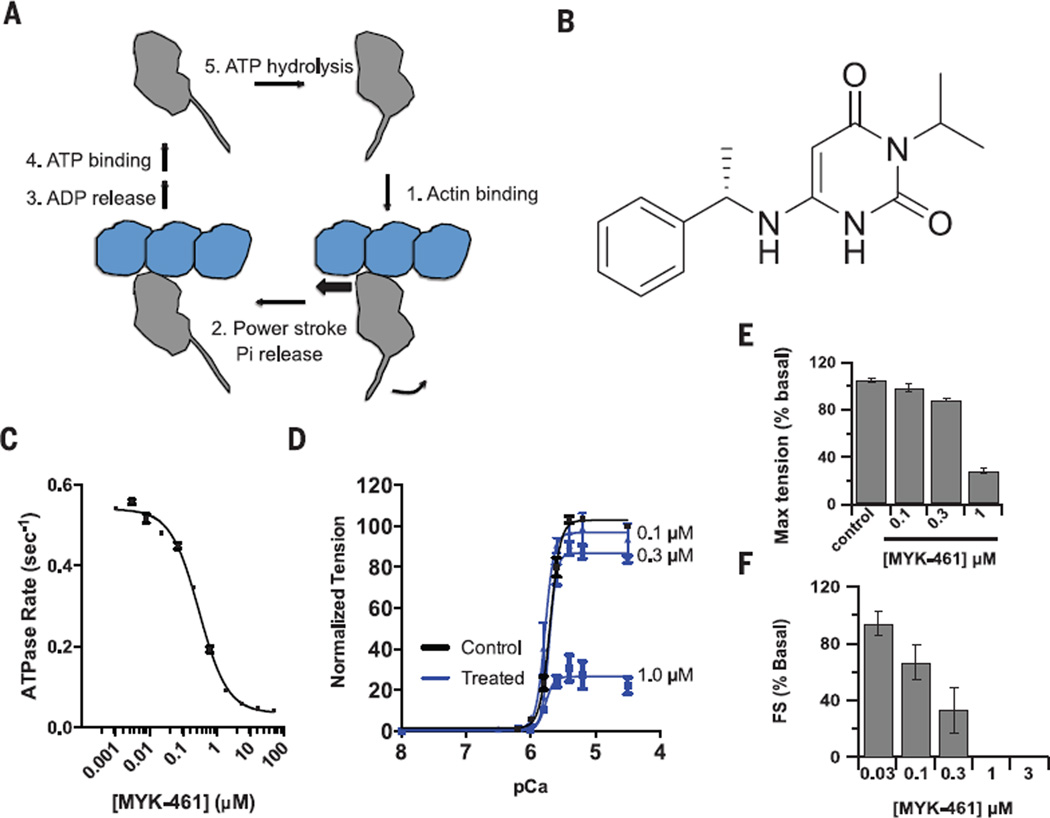
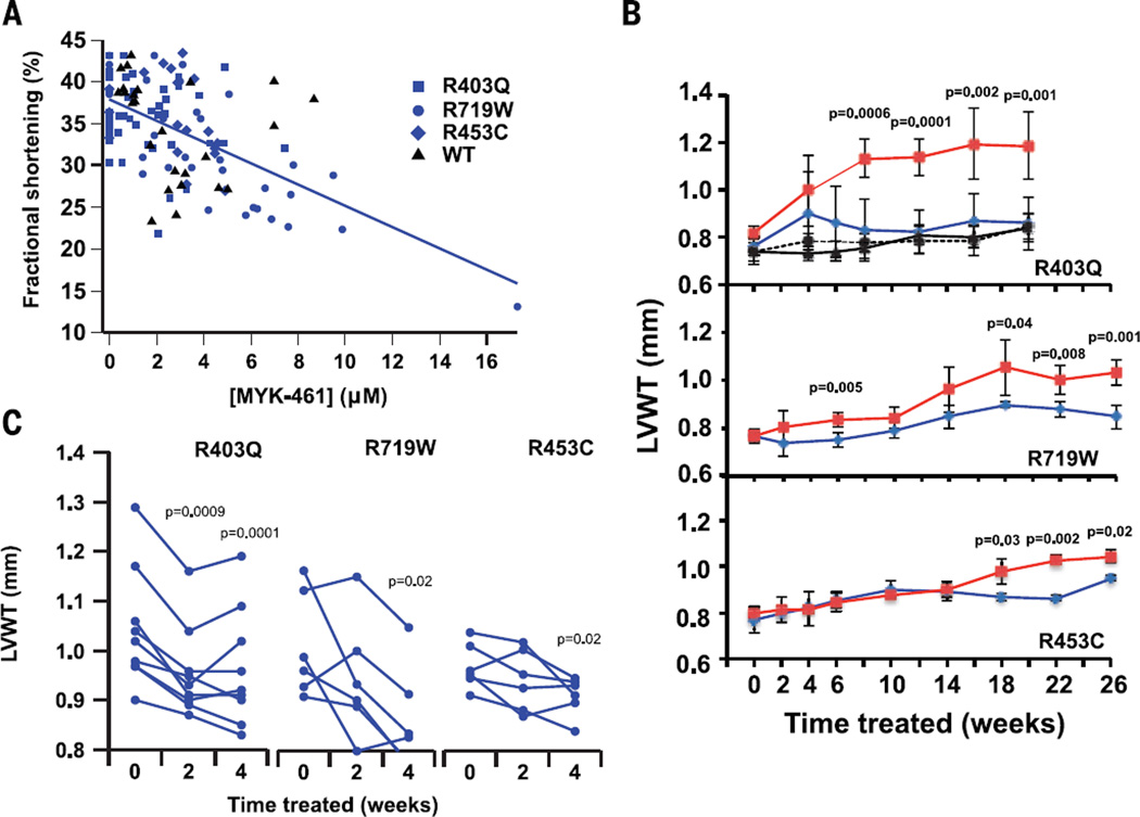
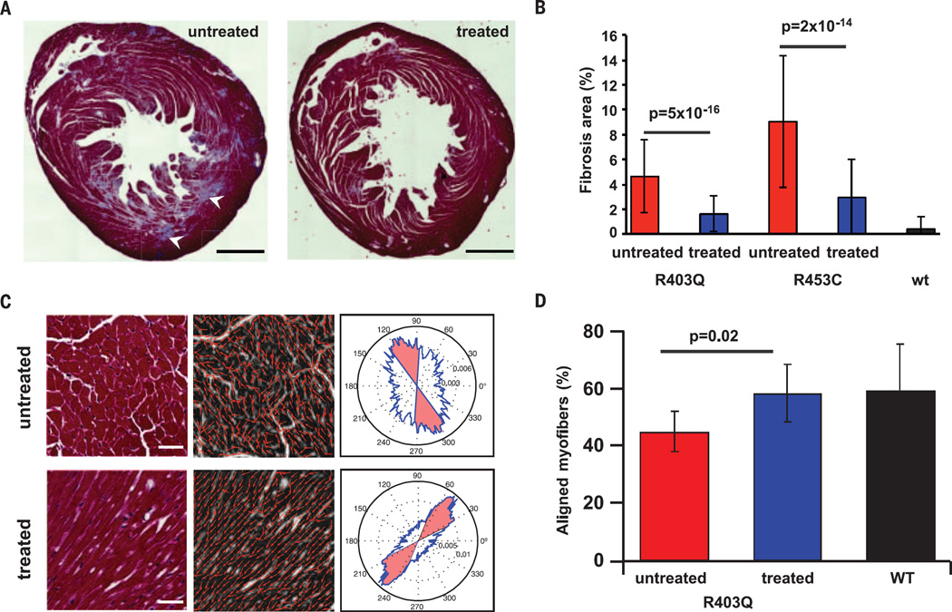
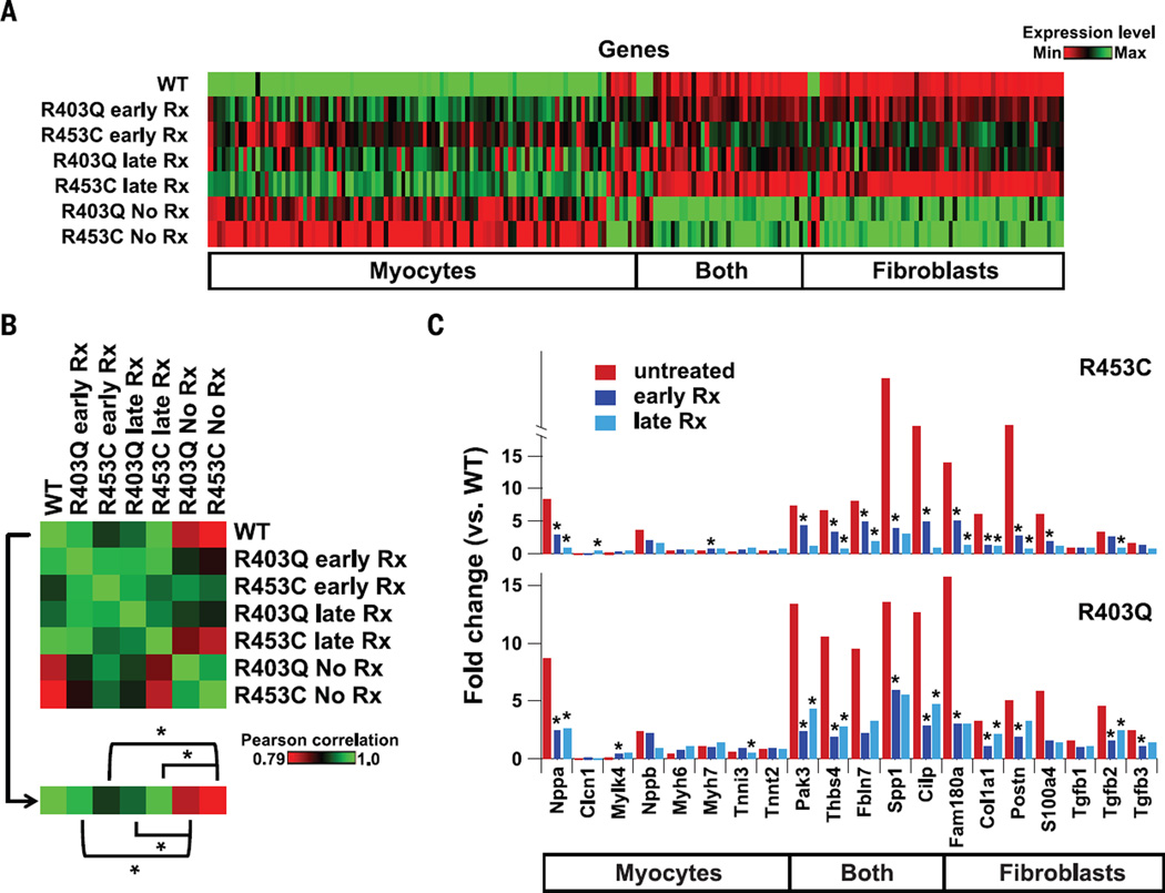
Hypertrophic cardiomyopathy (HCM) is an inherited disease of heart muscle that can be caused by mutations in sarcomere proteins. Clinical diagnosis depends on an abnormal thickening of the heart, but the earliest signs of disease are hyperdynamic contraction and impaired relaxation. Whereas some in vitro studies of power generation by mutant and wild-type sarcomere proteins are consistent with mutant sarcomeres exhibiting enhanced contractile power, others are not. We identified a small molecule, MYK-461, that reduces contractility by decreasing the adenosine triphosphatase activity of the cardiac myosin heavy chain. Here we demonstrate that early, chronic administration of MYK-461 suppresses the development of ventricular hypertrophy, cardiomyocyte disarray, and myocardial fibrosis and attenuates hypertrophic and profibrotic gene expression in mice harboring heterozygous human mutations in the myosin heavy chain. These data indicate that hyperdynamic contraction is essential for HCM pathobiology and that inhibitors of sarcomere contraction may be a valuable therapeutic approach for HCM.


Figures

Fig. 1
Fig. 1. MYK-461 inhibits myosin ATPase and contractility of cardiomyocytes
(A) Chemomechanical cycle of myosin, demonstrating the coupling between ATP hydrolysis and the myosin powerstroke. The swinging of the myosin lever arm (gray; bound light chains not shown) in step two is associated with movement of the actin filament (blue). (B) Chemical structure of MYK-461. (C) Decreasing rate of myosin ATPase in mouse cardiac myofibrils treated with increasing concentrations ofMYK-461 (n = 6 biological replicates, mean ± SD). (D) Plots of tension versus pCA for skinned cardiac muscle fibers (from rats) exposed to increasing concentrations of MYK-461 (n = 3 to 4 fibers per concentration, mean ± SEM). (E) Decreasing maximal tension of skinned cardiac muscle fibers (from rats) exposed to increasing concentrations of MYK-461 (n = 3 to 4 fibers per concentration, mean ± SEM). (F) Decreasing fractional shortening (FS) of isolated adult rat ventricular cardiomyocytes treated with increasing concentrations of MYK-461 (n = 3 to 5 cells per concentration, mean ± SD).
Fig. 2
Fig. 2. MYK-461 reduces cardiac contractility in mouse models of HCM and prevents or ameliorates LV hypertrophy
(A) Reduction in fractional shortening with increasing MYK-461 plasma exposure in hearts from R403Q, R719W, R453C, and WT mice, fit by linear regression (n = 6 to 9 mice per background, Pearson’s r = 0.57, P < 105). (B) Changes in LVWT over time by echocardiography for WT mice (top panel: dashed black line, untreated; solid black line, treated; n = 6 mice each) and the indicated HCM mouse models (blue, treated with MYK-461; red, untreated; n = 3 to 6 mice per background). All data are expressed as mean ± SD, and statistical significance was tested by repeated-measures one-way analysis of variance. (C) Reduction in LVWT for the indicated HCM mouse models treated with MYK-461 after the onset of hypertrophy (15 to 30 weeks of age). Statistical significance was tested by paired t tests compared with baseline values.
Fig. 3
Fig. 3. MYK-461 reduces the development of myocardial disarray and fibrosis in mouse models of HCM
(A) Sections from untreated (left) and MYK-461–treated (right) 30-week-old R403Q mice stained with Masson’s trichrome (arrowheads point to fibrotic areas). Scale bars, 1 mm. (B) Reduction in fibrosis area with MYK-461 treatment assessed in more than eight sections per mouse for R403Q and R453C HCM mice (n = 5 to 6 animals per group). (C) Representative regions of interest for analysis of cell orientation fromR403Q mouse heart sections. Regions are shown (left) stained with Masson’s trichrome (scale bars, 50 µM), (middle) with local gradient vectors illustrating cell orientations for analysis, and (right) as rose plots of the distribution of myofibril orientation angles (blue line; myofibrils oriented within 20° of the mean are shaded in red). (D) Percentage of aligned myofibers in WT and R403Q mice with and without MYK-461 treatment (n = 6 animals per condition). All data expressed are as mean ± SD; statistical comparisons were performed by t test.
Fig. 4
Fig. 4. MYK-461 suppresses hypertrophic and profibrotic gene programs in mouse models of HCM
(A) Heat map illustrating levels of gene expression in WT and treated and untreated HCM mice for 200 genes that are highly differentially regulated in HCM mice compared with WT mice. Genes are divided according to the cell type in which they are expressed (myocytes, fibroblasts, or both). Treatment with MYK-461 (2.5 mg/kg per day) began either before overt hypertrophy (early Rx) or after the development of hypertrophy (late Rx). (B) Heat map showing Pearson correlations between mouse genetic backgrounds and MYK-461 treatment regimens for 200 genes differentially regulated in untreated HCM mice. Statistical comparisons were performed between the Pearson correlations for WT animals or untreated mutants and each treatment condition for that mutant. Significance was assessed by Fisher r-to-z transformation (*P < 0.0001). (C) Fold change in gene expression levels (as compared with WT animals) for selected genes expressed in myocytes, fibroblasts, or both across MYK-461 treatment regimens in R453C mice (top) and R403Q mice (bottom; *P < 0.05).

Comment in

Similar articles

Cited by

References

    1. Maron BJ, et al. Circulation. 1995;92:785–789. - PubMed
    1. Klein MD, Lane FJ, Gorlin R. Am. J. Cardiol. 1965;15:773–781. - PubMed
    1. Wilson WS, Criley JM, Ross RS. Am. Heart J. 1967;73:4–16. - PubMed
    1. Stewart S, Mason DT, Braunwald E. Circulation. 1968;37:8–14. - PubMed
    1. Maron BJ, et al. JAMA. 1999;281:650–655. - PubMed

Publication types

MeSH terms